Description
Huvitz All –in-One OCT
All-in-One HOCT in smart 3D OCT & Fundus Camera, Totally integrated system combined with PC Provides OCT and Fundus data on one Screen. All-in-One HOCT is easy to use. One button, it creates a High-speed Scan and a High-Quality Image. Huvitz All-in-One HOCT will be the icon for leading a new era of Optical Coherence Tomography (OCT).
Salient Features:-
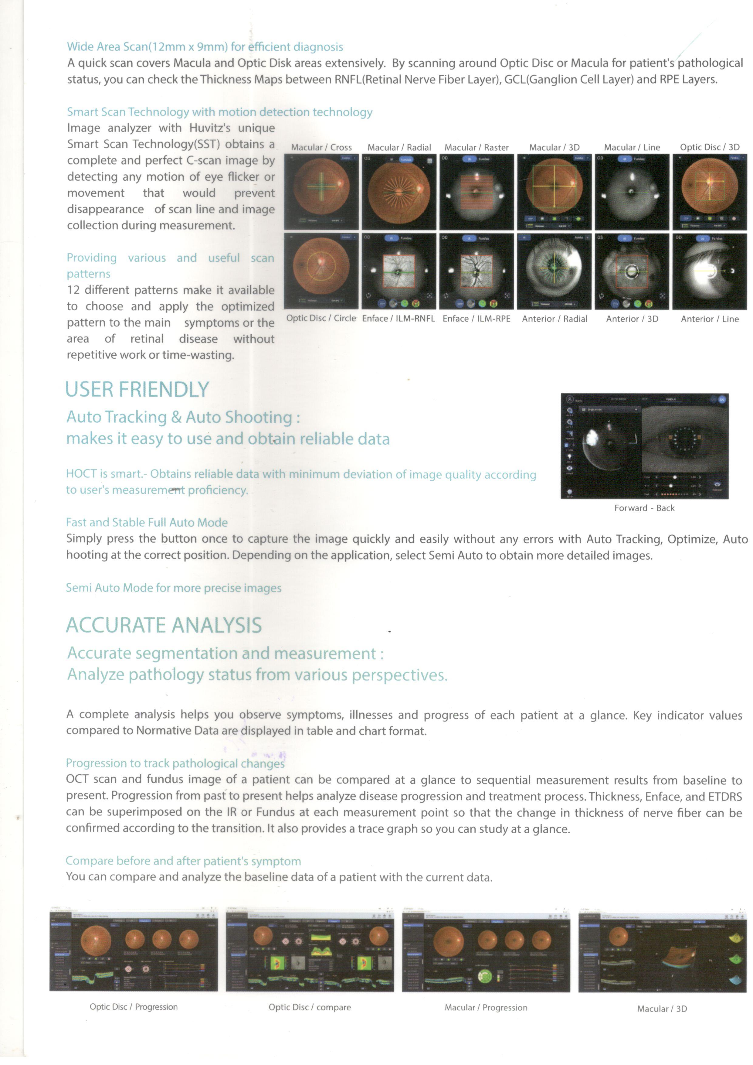
Wide Area Scan (12mm x 9mm) for efficient diagnosis
A quick scan covers Macula and Optic Disk areas extensively. By scanning around Optic Disc or Macula for patient’s pathological status, you can check the Thickness Maps between RNF (Retinal Nerve Fiber Layer), GCL (Ganglion Cell Layer) and RPF Layers.
Smart Scan Technology with motion detection technology
Providing various and useful scan patterns
USER FRIENDLY
Auto Tracking & Auto Shooting:
- Makes it easy to use and obtain reliable data
- HOCT is smart-Obtains reliable data with minimum deviation of image quality according to user’s measurement proficiency.
- Fast and Stable Full Auto Mode
- Semi Auto Mode for more precise images
ACCURATE ANALYSIS
Accurate segmentation & measurement:
- Analyze pathology status from various perspectives.
- Progression to track pathological changes
- Compare before and after patient’s symptom
ANTERIOR MEASUREMENT
One Single System:
- Start and finish in one place, making patient more comfortable.
- 9mm Wide Chamber View / Corneat Thickness
- Corneal Thickness Map
FULL COLOR FUNDUS IMAGE
Insight of Posterior Segment of Eye:
- Auto-Detection of Pupil Size and Auto Flash Level Function
- Panorama function for wide range of peripherals
DETAILED REPORT
From quick summary to simple comparison and complex evaluation:
- Complete a perfect report
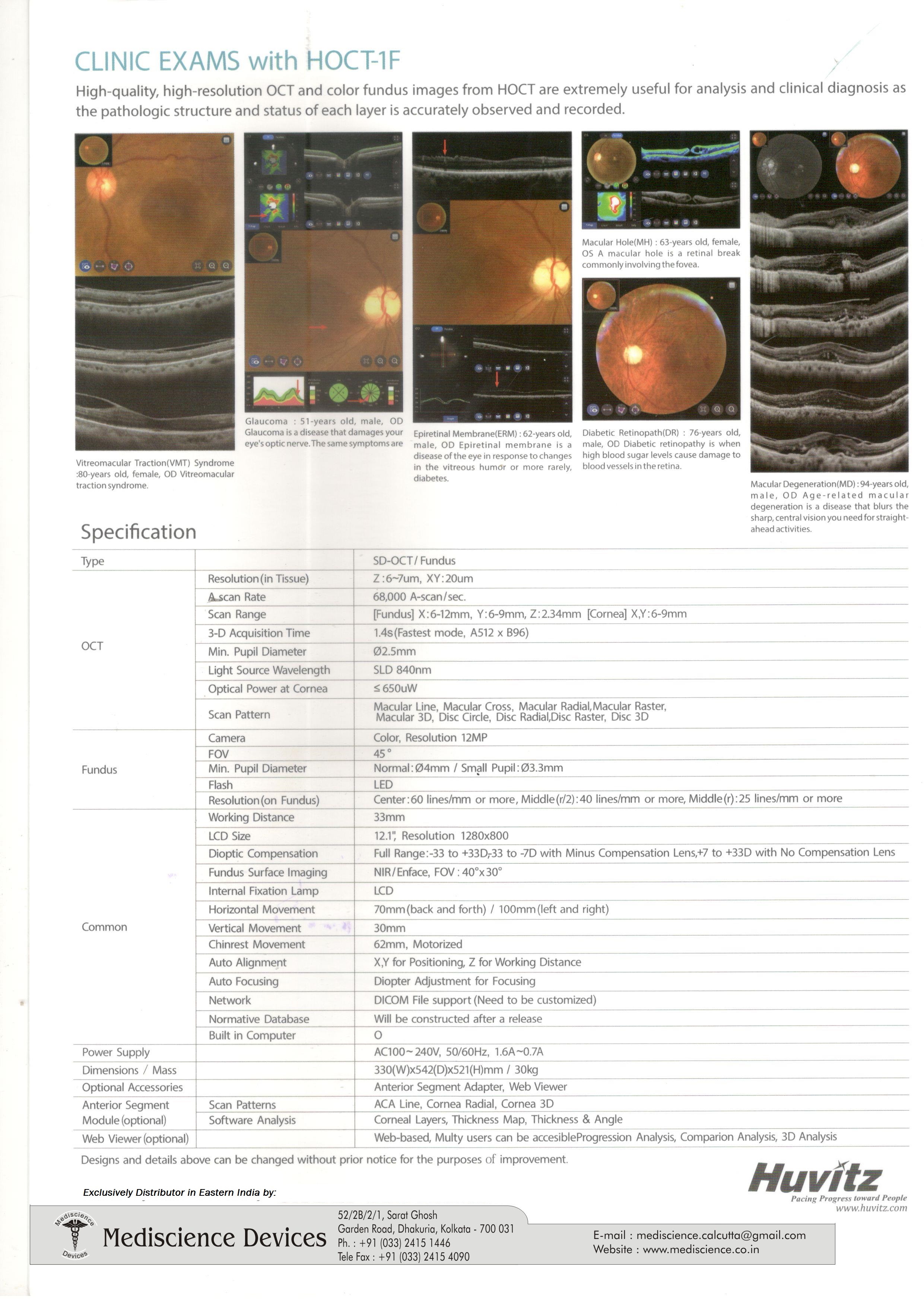
CLINIC EXAMS with HOCT-1F
High-quality, high-resolution OCT and color fundus images from HOCT are extremely useful for analysis and clinical diagnosis as the pathologic structure and status of each layer is accurately observed and recorded.


Reviews
There are no reviews yet.