The Ultimate Buyer’s Guide to Portable Eye Ultra Sound: Unlocking Clinical Precision with Advanced B Scan Technology
In modern ophthalmology, accurate visualization of the posterior segment of the eye is non-negotiable. When optical media is compromised by dense cataracts, corneal opacities, hyphema, or vitreous hemorrhages, traditional optical diagnostic tools—such as Optical Coherence Tomography (OCT) systems, slit lamp biomicroscopes, and fundus cameras—are rendered completely blind. In these critical clinical scenarios, high-frequency acoustic imaging becomes the clinician’s only diagnostic window.
Today, procuring a high-precision portable eye ultra sound system is not merely a luxury upgrade—it is a foundational requirement for any competitive eye hospital, trauma center, or multi-specialty OPD setup.
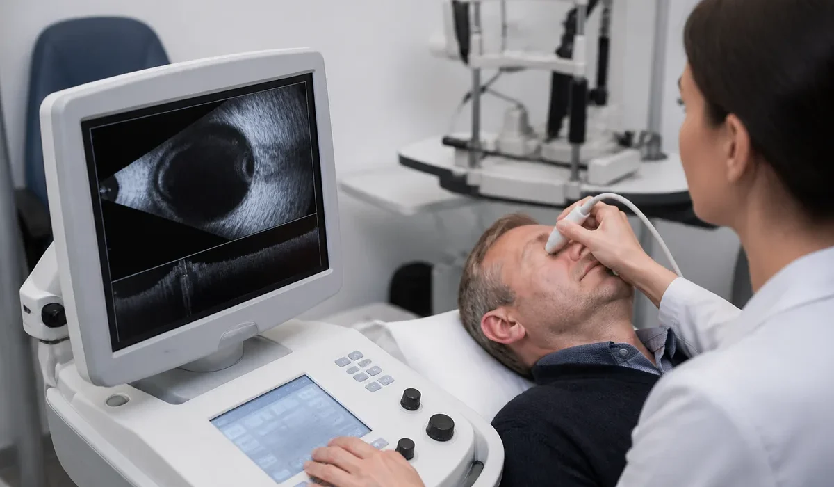
The rapid evolution of ocular sonography has shifted the market from heavy, tethered console machines to lightweight, highly mobile systems. Armed with a modern b scan machine, ophthalmologists can instantly evaluate retinal detachments, intraocular tumors, posterior vitreous detachments, and retrobulbar masses directly at the patient’s bedside or in the consultation chair.
As a premier medical equipment supplier with over 27 years of institutional dominance across Eastern and North-Eastern India, Mediscience Devices presents this enterprise-grade commercial buyer’s guide. In this deep dive, we evaluate acoustic physics, clinical applications, spatial workflows, and the total cost of ownership (TCO) for two of the most dominant B-scan technologies available today: the Matrix Fujitron Digital Pocket Wireless B-Scan (Model EU-1C) and the Suoer A/B Scan (Model SW-2000) workstation.
The Physics and Clinical Mandate of Ocular Ultrasound
Before allocating capital toward an ultrasound scanner, hospital procurement committees and clinical heads must evaluate the underlying physics and diagnostic scope of acoustic imaging.
High-Frequency Acoustic Transduction
Ophthalmic sonography utilizes acoustic frequencies significantly higher than general abdominal or obstetric sonography—typically ranging between 8 MHz and 20 MHz.
Higher Frequency (11 MHz – 20 MHz): Provides exceptional axial and lateral resolution, allowing clinicians to resolve microscopic structural changes in the retina, macula, and vitreoretinal interface.
Lower Frequency (8 MHz – 10 MHz): Provides deeper acoustic penetration, necessary for pushing past the posterior sclera to evaluate deep orbital lesions, retrobulbar hemorrhages, and extraocular muscle thickening.
Resolving Diagnostic Ambiguity: A-Scan vs. B-Scan
A-Scan (Amplitude Scan): A one-dimensional acoustic scan that measures the time delay of echoes along a single linear path. It converts echo amplitudes into vertical spikes on a baseline, primarily used to measure the eye’s axial length for Intraocular Lens (IOL) power calculation prior to cataract surgery.
B-Scan (Brightness Scan): A two-dimensional b mode ultrasound scan produced by sweeping an acoustic beam across the globe. Returning echoes are converted into brightness-modulated dots on a screen, forming a high-definition grayscale image of the structural anatomy within the eye and orbit.
Combining an a scan b scan protocol ensures that a clinic can handle both pre-operative optical biometry and complex posterior segment pathology screening under one roof.
Strategic Workstation vs. Pocket Wireless B-Scan: A Commercial Comparison
When preparing to buy portable ultrasound technology, hospital directors face a choice between two distinct form factors: ultra-mobile wireless probes and complete desktop workstations. Understanding how these systems fit into your clinical workflow is essential for maximizing diagnostic throughput and financial ROI.
Comparative Analysis Matrix
| Evaluation Parameter | Matrix Fujitron EU-1C (Wireless) | Suoer A/B Scan SW-2000 (Workstation) |
| Form Factor | Handheld wireless b scan probe (<150g) | Desktop module with dedicated laptop & printer |
| Acoustic Frequencies | Selectable Dual-Frequency (8 MHz & 11 MHz) | High-Resolution 10 MHz B-Scan Transducer |
| Display Ecosystem | Open BYOD (iOS, Android, Windows Tablets) | Pre-configured, high-definition hospital laptop |
| Data Transmission | Zero-latency Wi-Fi hotspot connection | USB / Direct cable interface |
| Power Source | Internal rechargeable Li-ion battery | AC Mains powered workstation |
| Reporting & Output | Digital export, DICOM, instant image share | Integrated thermal/inkjet paper report printer |
| Target Clinical Zone | Emergency rooms, ICUs, outreach camps, mobile OPD | Dedicated imaging suites, cataract OPD stations |
Product Deep Dive 1: Matrix Fujitron Digital Pocket Wireless B-Scan (Model EU-1C)
For healthcare facilities seeking maximum mobility, seamless teleophthalmology integration, and modern ergonomics, the Matrix Fujitron Digital Pocket Wireless B-Scan (Model EU-1C) represents the pinnacle of modern portable eye ultra sound technology.
1. Zero-Cable Ergonomics & Solid-State Engineering
Traditional wired probes suffer from cable drag, which exerts subtle tension on the physician’s hand during delicate kinetic scans. The Fujitron EU-1C houses its entire acoustic engine, power supply, and wireless transmitter inside a solid-state probe weighing less than 150 grams.
Preventing Strain: Eliminates physical cable drag, allowing smooth, uninhibited probe angulation across all four quadrants of the eye.
Infection Control: Devoid of thick, ribbed cables that drag across clinical surfaces, the smooth, water-resistant casing can be disinfected between patients in seconds with standard medical-grade wipes.
2. Dual-Frequency Selectivity: 11 MHz vs. 8 MHz
The EU-1C is one of the few portable devices offering selectable dual-frequency modes:
11 MHz High-Resolution Mode: Provides crisp 256-grayscale resolution for evaluating subtle macular holes, retinal detachment margins, and vitreous membrane traction.
8 MHz Deep-Penetration Mode: Pushes past the sclera to evaluate extraocular muscle bellies, retrobulbar optic nerve sheath diameter (ONSD), and deep orbital masses.
3. Open BYOD (Bring Your Own Device) Ecosystem
One of the most predatory commercial practices in medical technology is vendor-lock-in—forcing hospitals to purchase expensive, proprietary display monitors. The Fujitron EU-1C operates on an open platform:
Hardware Agnostic: Connects via its self-generated Wi-Fi hotspot to any commercial iPad, Android tablet, or Windows Surface.
Zero Subscription Fees: The high-definition diagnostic software is free to install across multiple devices. If a tablet breaks in your clinic, simply install the app onto another tablet and resume scanning immediately—eliminating vendor lock-in and expensive monitor replacement costs.
4. Continuous Kinetic Scanning & Teleophthalmology Export
The EU-1C captures smooth, real-time cine-loops (video sequences) of the eye during kinetic movement. Clinicians can capture dynamic scans, freeze frames, perform distance/area measurements, and export full diagnostic packages instantly via Wi-Fi or cellular networks for immediate teleophthalmology consultations.
Product Deep Dive 2: Suoer A/B Scan Model SW-2000 (Complete Turnkey Workstation)
While wireless probes excel in point-of-care mobility, high-volume cataract hospitals and centralized diagnostic rooms often prefer a stationary, turnkey station. The Suoer A/B Scan Model SW-2000 provides a robust, all-in-one workstation engineered for high-volume clinical throughput.
1. Complete Turnkey Procurement Bundle
Hospital procurement departments frequently encounter driver incompatibilities and software installation failures when trying to pair diagnostic probes with commercial PCs. The Suoer SW-2000 package supplied by Mediscience eliminates IT friction entirely:
Pre-Configured Hardware: Arrives complete with the central processing module, high-resolution b scan probe, an industry-branded laptop pre-loaded with diagnostic software, and an integrated printer.
Plug-and-Play Setup: Ready for immediate clinical deployment straight out of the crate.
2. Clinical Precision & Grayscale Fidelity
The SW-2000 utilizes a 10 MHz sector-scanning probe that delivers exceptional acoustic detail across a 53-degree scanning angle:
256-Level Grayscale Resolution: Ensures subtle variations in acoustic tissue density are clearly visible.
Time Gain Compensation (TGC): Features adjustable near, far, and overall gain controls, enabling clinicians to boost weak echoes from posterior vitreous membranes without over-saturating strong scleral signals.
3. Comprehensive Reporting & Expandable Architecture
Instant Hard-Copy Output: The included high-speed printer allows clinicians to generate color or grayscale diagnostic reports immediately following the examination. Hard-copy documentation can be handed directly to the patient or filed in their physical medical chart.
Expandable A-Scan Capability: While configured with the high-resolution B-scan probe, the SW-2000 console features dual-port architecture that supports add-on A-scan biometry probes, turning the unit into a complete biometry and diagnostic station for cataract surgery planning.
Clinical Application Breakdown: Diagnostic Scenarios
A high-performance b scan machine is essential across a wide spectrum of ocular pathologies. Below are key clinical scenarios where sonography provides decisive diagnostic evidence:
1. Differentiating Retinal Detachment (RD) from Posterior Vitreous Detachment (PVD)
When dense vitreous hemorrhage obscures the retina, sonography is the only way to rule out an underlying retinal tear:
Rhegmatogenous Retinal Detachment: Appears as a continuous, highly reflective, thick membrane anchored firmly to the optic disc and the ora serrata. On kinetic scanning, it exhibits limited mobility.
Posterior Vitreous Detachment: Appears as a thin, low-to-medium reflective membrane that sways freely on eye movement (“after-movement”) and is not anchored to the optic nerve head.
2. Ocular Trauma & Foreign Body Localization
In penetrating ocular injuries, visual inspection is often impossible due to hyphema or corneal laceration:
Intraocular Foreign Bodies (IOFB): Metallic, glass, or stone fragments produce brilliant, extremely high-amplitude echoes accompanied by characteristic trailing “comet-tail” or acoustic shadowing artifacts.
Globe Rupture Verification: Sonography maps the integrity of the posterior scleral wall, identifying occult posterior scleral tears without touching the open wound directly.
3. Ocular Oncology & Choroidal Mass Evaluation
When a pigmented mass is noted in the fundus, a b scan ultrasonography exam quantifies lesion parameters:
Choroidal Melanoma: Typically demonstrates a classic smooth, dome-shaped or mushroom-shaped elevation, internal acoustic hollowness (low-to-medium internal reflectivity), and choroidal excavation.
Choroidal Hemangioma: Exhibits high uniform internal reflectivity without acoustic hollowness, helping differentiate benign vascular tumors from malignant lesions.
Procurement Checklist for Hospital Administrators
Before issuing purchase orders for a portable ultrasound machine, hospital procurement committees should evaluate prospective vendors against this operational checklist:
Regulatory & Quality Standards
Regulatory Clearance: Is the device CDSCO-registered under Indian Medical Device Regulations?
Optical/Acoustic Standards: Does the unit carry valid CE or ISO 13485 (Medical Devices) manufacturing certifications?
Hardware & Software Performance
Frequency Selectivity: Does the device provide appropriate acoustic frequencies (8 MHz – 11 MHz for B-scan; 10 MHz for A-scan biometry)?
Display & Connectivity: Does the software support open digital export (JPG, PNG, DICOM) without hidden annual licensing fees?
Service Infrastructure & Long-Term SLA
Local Spare Parts Inventory: Does the distributor maintain local stocks of replacement transducers, cables, and circuit boards?
Uptime SLA: Does the contract guarantee technician dispatch within 24 to 48 hours to prevent OPD disruption?
The Mediscience Advantage: 27+ Years of Institutional Trust
Purchasing complex ophthalmic equipment is a decade-long commitment. Buying imported hardware from unverified drop-shippers or generic online vendors exposes your facility to severe operational risks—including lack of local calibration, missing replacement parts, and extended downtime.
Mediscience Devices has served as the backbone of eye care infrastructure across Eastern and North-Eastern India for over 27 years. Headquartered in Kolkata, our institutional reach spans West Bengal, Jharkhand, Odisha, Bihar, Chhattisgarh, Assam, Nagaland, Mizoram, Manipur, and Tripura.
When you procure your eye ultrasound equipment through Mediscience Devices, you secure:
Transparent Pricing: Factory-direct pricing that eliminates middleman inflation and unnecessary corporate advertising markups.
Turnkey Technical Delivery: Doorstep installation and precision acoustic calibration performed by factory-trained biomedical engineers.
Comprehensive Application Training: On-site clinical training for your doctors, optometrists, and sonography technicians to ensure your staff maximizes the software’s diagnostic capabilities.
Zero-Downtime Guarantee: Local inventory of spare transducers, probes, and loaner units to keep your clinic fully operational while any service is performed.
Conclusion: Elevate Your Diagnostic Capability Today
A high-performance portable eye ultra sound system is an indispensable asset that elevates diagnostic accuracy, prevents unnecessary patient referrals, and drives surgical volume.
Whether your facility requires the extreme mobility and open tablet ecosystem of the Matrix Fujitron EU-1C Wireless B-Scan or the complete, turnkey reporting setup of the Suoer A/B Scan SW-2000 Workstation, Mediscience Devices provides the authentic technology, transparent pricing, and local engineering support you need.
Take the next step in modernising your posterior segment diagnostics. Contact the B2B Procurement Experts at Mediscience Devices today to request a commercial quote, confirm product availability, or schedule an on-site clinical demonstration.
People Also Ask - Commercial & Clinical FAQs
An A-scan (Amplitude scan) is a one-dimensional acoustic measurement used primarily to determine the axial length of the eye for calculating Intraocular Lens (IOL) power before cataract surgery. A B-scan (Brightness scan) provides a two-dimensional, cross-sectional grayscale image of the eye, allowing doctors to evaluate structural pathologies like retinal detachments, vitreous traction, and tumors.
No. The Matrix Fujitron EU-1C operates on an open "Bring Your Own Device" (BYOD) framework. It connects via zero-latency Wi-Fi to any standard commercial iOS (iPad), Android, or Windows tablet, saving clinics the substantial cost of purchasing and maintaining proprietary medical monitors.
Mediscience Devices, located in Kolkata, is a premier authorized supplier of top-tier ophthalmic and optometry equipment. Serving Eastern and North-Eastern India for over 27 years, they offer full installation, clinical staff training, transparent pricing, and rapid on-site technical support for all ocular ultrasound systems.
The Suoer A/B Scan Model SW-2000 package supplied by Mediscience Devices is a complete turnkey workstation. It includes the central diagnostic module, a high-resolution 10 MHz B-scan probe, an industry-branded compatible laptop pre-loaded with diagnostic software, and an integrated printer for immediate report generation.