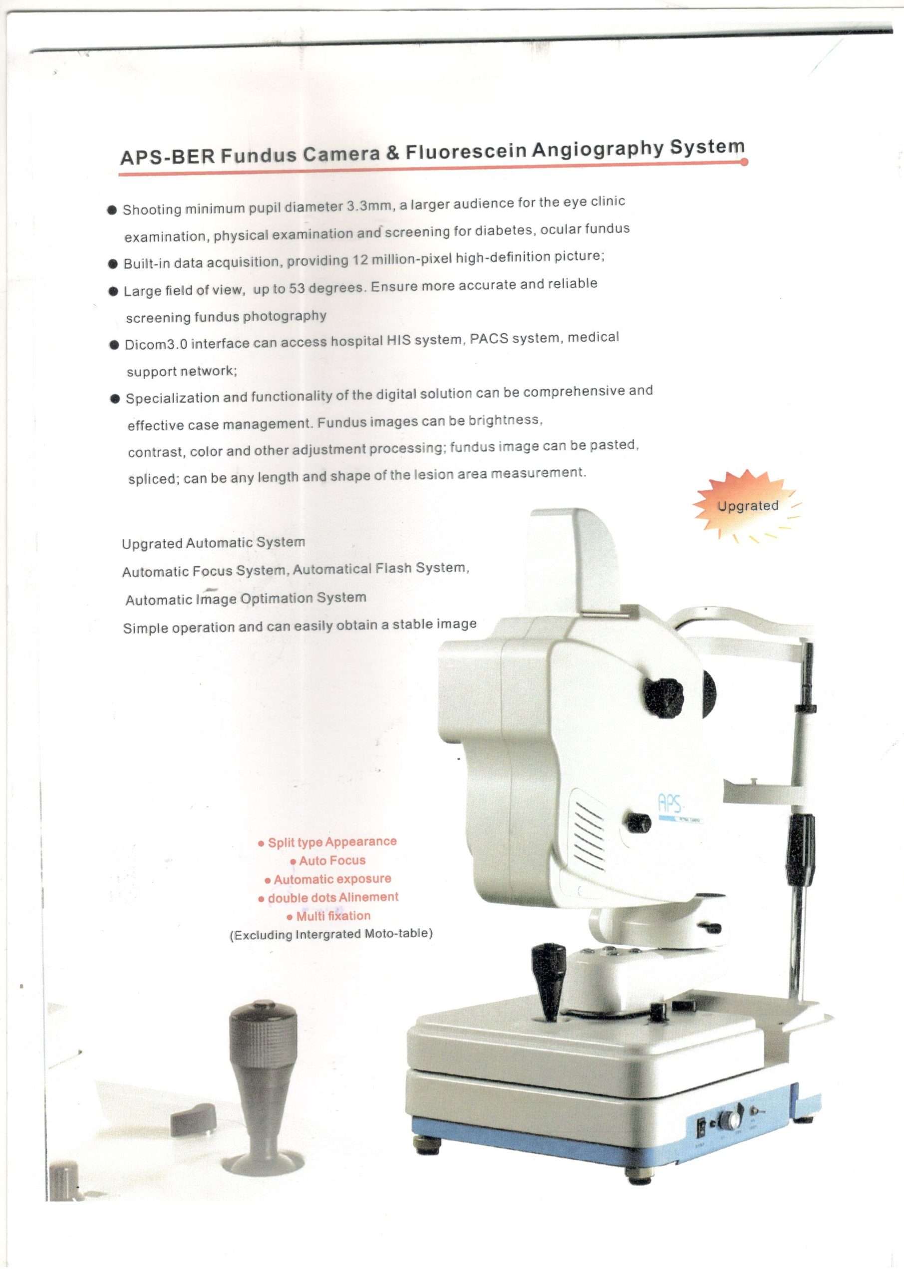
APS -BER Fundus Camera & Fluorescein Angiography System

*Shooting minimum pupil diameter 3.3mm, a larger audience for the eye clinic examination, physical examination and screening for diabetes, ocular fundus; *Built-in data acquisition, providing 12 million-pixel high-defination picture; * Large field of view, up to 53 degrees. Ensure more accurate and reliable screening fundus photography;  * Dicom 3.0 interface can access hospital HIS system, PACS system, medical support network; * Specialization and functionality of the digital solution can be comprehensive and effective case management. Fundus images can be brightness, contrast, color and other adjustment processing; fundus image can be pasted, spliced; can be any length and shape of the lesion area measurement.

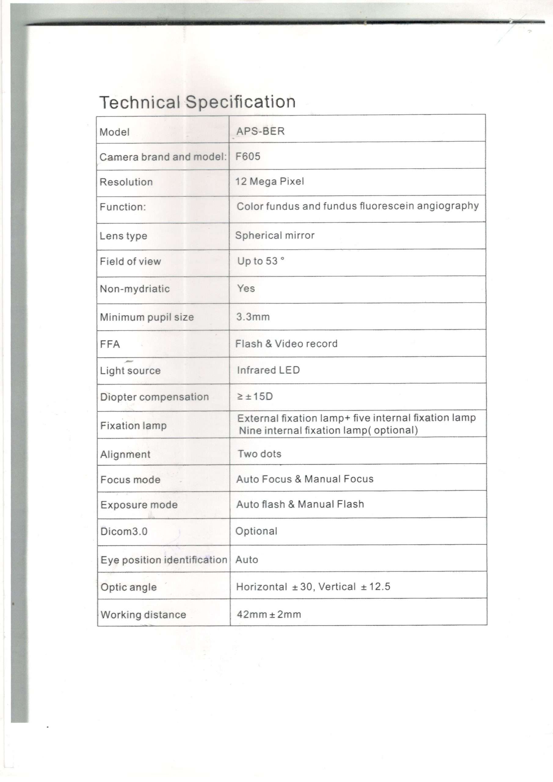
TECHNICAL SPECIFICATIONS:-

Camera Resolution 12 Mega Pixel
Function Color fundus and fundus fluoresce in angiography
Lens type Spherical mirror
Field of view Up to 53  ̊
Non-mydriatic Yes
Minimum pupil size 3.3mm
FFA Flash & Video Record
Light source Infrared LED
Diopter compensation ≥ ± 15D
Fixation lamp External fixation lamp+ five internal fixation lamp Nine internal fixation lamp (optional)
Alignment Two dots
Focus mode Auto Focus & Manual Focus
Exposure mode Auto flash & Manual Flash
Dicom3.0 Optional
Eye position identification Auto
Optic angle Horizontal ± 30, Vertical ± 12.5
Working distance 42mm ± 2mm

Download Catalogue

http://www.mediscience.co.in/wp-content/uploads/2018/02/Kangua-Fundus-Camera-APS-BER-2.pdf

Category:

Reviews

There are no reviews yet.

Be the first to review “APS -BER Fundus Camera & Fluorescein Angiography System”

Your email address will not be published. Required fields are marked *