
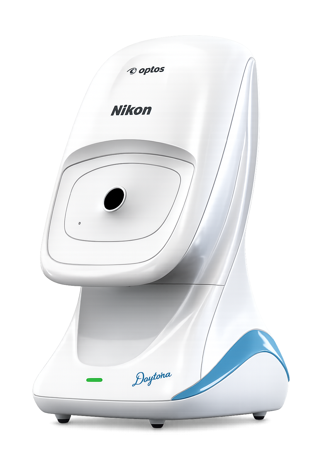

Description
Optos Daytona Ultra-Widefield Fundus Camera for Retinal Imaging & Ophthalmology
The Optos Daytona Ultra-Widefield Fundus Camera is an advanced retinal imaging platform designed to expand the scope of routine fundus examination beyond the central retina. Built around Optos’ proprietary optomap® technology, the system provides 200° ultra-widefield retinal imaging, enabling clinicians to capture approximately 82% of the retina in a single image.
Unlike conventional fundus imaging systems that primarily focus on the posterior pole, the Optos Daytona is designed to provide a much broader view extending toward the retinal periphery. This makes it a valuable retinal camera for screening, documentation and clinical assessment where peripheral retinal visibility can be important.
The system combines high-speed, non-contact and non-mydriatic imaging with multiple image modalities, helping ophthalmologists and optometrists integrate retinal imaging into routine clinical workflows. With image acquisition in less than 0.4 seconds, Daytona can support efficient patient throughput while providing a comfortable examination experience.
Advanced Widefield Retinal Imaging in a Single Capture
One of the key advantages of the Optos fundus camera platform is its ability to capture a much larger area of the retina than conventional fundus cameras. The Daytona’s 200° field of view allows approximately 82% of the retina to be visualised in one capture, supporting a more comprehensive retinal examination.
This wide field retinal imaging capability can be particularly useful when documenting peripheral retinal findings, monitoring retinal disease and assessing patients who require broader retinal visualisation. The system is also designed to produce high-quality images through pupils as small as 2 mm and through most cataracts, according to the supplied product information.
Non-Mydriatic, Non-Contact Retinal Imaging
The Daytona combines the advantages of a non mydriatic fundus camera with ultra-widefield imaging. In most patients, retinal images can be acquired without pharmacological pupil dilation, helping streamline the examination process and reduce additional preparation time.
Its non-contact design also avoids direct corneal contact during image acquisition, creating a more comfortable experience for patients and making the system suitable for repeated retinal imaging and screening workflows.
Multiple Imaging Views for Comprehensive Retinal Assessment
The Optos Daytona is not limited to conventional colour retinal photography. Its imaging capabilities include several complementary views that can provide additional information during retinal assessment:
- Colour imaging for general retinal documentation
- Red-free / Sensory View for enhanced visualisation of retinal structures
- Choroidal View for choroidal imaging
- Green autofluorescence for autofluorescence-based imaging
- Stereo optic disc imaging for optic nerve assessment
This combination allows the retinal camera to be incorporated into different ophthalmic examination and documentation workflows rather than functioning solely as a conventional fundus photography system.
Designed for Efficient Ophthalmic Workflows
Speed is another important consideration for busy eye-care facilities. Daytona captures diagnostic-quality retinal images in less than 0.4 seconds, helping reduce examination time and supporting higher patient throughput.
The system works alongside OptosAdvance™, a browser-based image management platform that provides tools for image comparison, measurements, referral management and electronic image handling. The supplied specifications also identify DICOM compatibility, making the system suitable for practices looking to integrate retinal imaging into a broader digital ophthalmology environment.
The software supports auto-montage up to 220°, extending the imaging capability when a wider composite retinal view is required.
Clinical Applications
The broad imaging capability of the Optos widefield platform makes Daytona suitable for a range of retinal and general ophthalmic workflows, including:
- Diabetic retinopathy screening
- Glaucoma evaluation
- Age-related macular degeneration assessment
- Peripheral retinal lesion documentation
- Retinal tear and detachment assessment
- Hypertensive retinopathy evaluation
- Routine retinal screening
- General ophthalmology
- Teleophthalmology and remote image review
- Retinal image documentation and follow-up
For high-volume eye-care environments, the combination of rapid acquisition, non-mydriatic imaging and wide retinal coverage can make ultra widefield retinal imaging a useful addition to the diagnostic workflow.
Technical Specifications
| Specification | Details |
|---|---|
| Imaging Field | 200° Ultra-Widefield |
| Retinal Coverage | Approximately 82% of retina in a single capture |
| Resolution | 20 μm (optomap), 14 μm (optomap Plus) |
| Imaging Modes | Colour, Red-Free, Choroidal, Green Autofluorescence |
| Exposure Time | Less than 0.4 seconds |
| Laser Wavelengths | 635 nm, 532 nm |
| Imaging Type | Non-Contact, Non-Mydriatic |
| Software | OptosAdvance™ |
| Communication | DICOM Compatible |
| Minimum Pupil | As small as 2 mm, according to supplied information |
Why Choose Optos Daytona from Mediscience?
For hospitals, retinal centres and high-volume ophthalmology practices, the Optos Daytona Ultra-Widefield Fundus Camera offers a combination of broad retinal coverage, rapid acquisition and multiple imaging modalities in a single retinal imaging platform.
Mediscience provides the Optos Daytona for professional ophthalmic facilities looking to implement advanced fundus camera, retinal camera and ultra-widefield retinal imaging capabilities. Our team can assist with product selection, commercial quotations, installation requirements, application support and after-sales service.
Looking to upgrade your retinal imaging workflow? Contact Mediscience for pricing, availability and B2B procurement assistance for the Optos Daytona Ultra-Widefield Fundus Camera.
Frequently Asked Questions
The Optos Daytona is a retinal imaging system used to capture widefield images of the retina for screening, documentation, monitoring and clinical assessment across a range of ophthalmic conditions.
The main advantage is its ultra-widefield retinal imaging capability. Daytona can capture a 200° field of view, allowing approximately 82% of the retina to be visualised in a single capture rather than focusing primarily on the central retina.
Yes. Its widefield imaging technology is designed to provide substantially broader retinal coverage, including areas extending toward the peripheral retina. This can be useful when assessing or documenting peripheral retinal findings.
Yes. Its wide retinal field makes it suitable for retinal screening workflows, including diabetic retinopathy screening and documentation.