Suoer AB-Scan SW-2000 Review: Elevating Diagnostic Accuracy in Cataract and Vitreoretinal OPDs
Modern ophthalmic practice relies heavily on advanced optical modalities such as Optical Coherence Tomography (OCT), slit-lamp biomicroscopy, and non-contact optical biometers. However, when an Outpatient Department (OPD) presents patients with opaque ocular media—such as mature dense cataracts, corneal leucomas, or acute vitreous hemorrhages—light-based optical equipment encounters total attenuation. In these critical clinical scenarios, optical diagnostics fail to yield diagnostic data or accurate axial measurements.
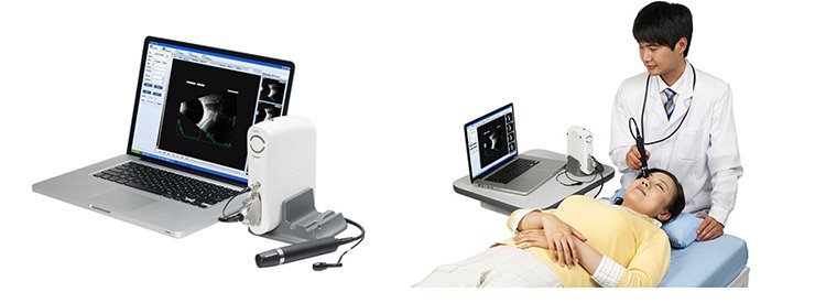
High-resolution ocular ultrasonography remains the gold-standard diagnostic foundation when light cannot penetrate the eye. The Suoer AB-Scan (Model SW-2000) provides a robust, laptop-integrated solution designed to bridge this exact diagnostic gap. Combining high-precision A-scan biometry with a 10 MHz sector B-scan imaging scanner, the Suoer SW-2000 offers surgical and vitreoretinal OPDs a portable, workstation-grade diagnostic tool.
This technical review explores the acoustic physics, clinical biometry calculations, posterior segment structural imaging capabilities, and operational advantages of integrating the Suoer AB-Scan into modern ophthalmic practices.
Ultrasonic Physics & Transducer Engineering in Ocular Diagnostics
Ocular ultrasound relies on the reflection of high-frequency sound waves across interfaces with differing acoustic impedances. In the eye, distinct boundaries exist between the cornea, aqueous humor, anterior/posterior lens capsules, vitreous humor, and retinal-choroidal-scleral complex.
The Suoer SW-2000 leverages specialized 10 MHz piezoelectric transducer technology across both its A-scan and B-scan modalities to deliver high spatial resolution while maintaining appropriate acoustic penetration through dense media.
Acoustic Performance Breakdown:
A-Scan Probe Mechanics: The a scan probe features a 10 MHz solid internal red-light fixation light source that aligns the acoustic axis with the patient’s visual axis. The signal processing engine allows for smooth dynamic gain adjustments (from 1 to 105 dB), enabling clinicians to amplify faint vitreous spikes without causing baseline electronic noise or distortion.
B-Scan Probe Mechanics: The sector-scanning b scan probe operates at a 10 MHz acoustic frequency, balancing axial resolution ($\le 0.2\text{ mm}$) with lateral resolution ($\le 0.4\text{ mm}$). It offers a wide scanning sector angle ($53^\circ$ to $60^\circ$), capturing extensive two-dimensional cross-sections of the posterior segment.
Gray-Scale Processing: The ultrasonic instrument processes acoustic reflections through 256 gray-scale levels, allowing subtle variations in vitreous echo density (such as distinguishing early vitreous haze from organized membranes) to be clearly visualized on screen.
Precision A-Scan Biometry: IOL Power Calculations & Formula Integration
In cataract surgery, achieving target refractive outcomes depends directly on the accuracy of pre-operative axial length (AL) measurements. An error of just $0.1\text{ mm}$ in axial length measurement leads to an approximate $0.25\text{ D}$ refractive error in the post-operative intraocular lens (IOL) calculation. When dense nuclear cataracts prevent optical biometers from acquiring signal spikes, a dedicated a scan instrument is essential.
The A-scan module in the Suoer AB-Scan provides automated, high-precision acoustic distance logging across all primary ocular media spaces.
| Anatomical Parameter | Measurement Capability | Clinical Significance |
| Axial Length (AL) | $15.0\text{ mm} – 40.0\text{ mm}$ (Accuracy: $\le \pm 0.05\text{ mm}$) | Primary variable in total IOL power determination |
| Anterior Chamber Depth (ACD) | $1.8\text{ mm} – 6.0\text{ mm}$ | Determines effective lens position (ELP) in 4th-gen formulas |
| Lens Thickness (LT) | $2.0\text{ mm} – 6.0\text{ mm}$ | Critical for phakic/cataractous lens volume evaluation |
| Vitreous Length (VL) | $10.0\text{ mm} – 30.0\text{ mm}$ | Evaluates posterior cavity dimensions |
Integrated Biometry Formulas & Velocity Compensation:
Different ocular tissues propagate acoustic waves at different velocities. The Suoer SW-2000 automatically compensates for tissue-specific acoustic velocities:
The onboard software integrates third- and fourth-generation IOL calculation algorithms, including SRK-II, SRK-T, HOLLADAY, HOFFER-Q, and HAIGIS, alongside dedicated post-refractive surgery calculation matrices. With auto-averaging features across multiple acquisition runs, the unit filters out anomalous velocity spikes, giving surgeons high confidence in a scan b scan ophthalmology planning.
High-Resolution B-Scan Imaging: Visualizing Hidden Posterior Segment Pathology
When visual axis opacities prohibit direct fundus visualization via ophthalmoscopy, a structural b scan becomes the definitive non-invasive diagnostic tool. The ophthalmic b scan capability of the SW-2000 converts high-frequency acoustic reflections into real-time, two-dimensional cross-sectional maps of the vitreous cavity, retina, choroid, and optic nerve head.
Key Clinical Diagnostic Applications:
Vitreous Pathology Assessment:
Vitreous Hemorrhage: Differentiates between fresh, low-amplitude dot-like echoes and dense, organized posterior vitreal clots.
Posterior Vitreous Detachment (PVD): Clearly delineates smooth, highly mobile posterior hyaloid membranes from fixed retinal structures.
Asteroid Hyalosis: Identifies classic, highly reflective, localized hyper-echoic calcium-lipid suspensions that glisten during dynamic eye movements.
Retinal & Choroidal Architecture:
Retinal Detachment (RD): Visualizes smooth, continuous, highly reflective membranes anchored at the optic disc and anterior ora serrata. Dynamic kinetic scanning helps differentiate rhegmatogenous RD from tractional or exudative subretinal fluid accumulation.
Choroidal Detachment: Displays smooth, dome-shaped, non-conical thick membranes that do not terminate at the optic disc.
Posterior Staphyloma: Maps localized outpouchings of the sclera in high myopes.
Ocular Trauma & Intraocular Lesions:
Intraocular Foreign Bodies (IOFBs): Pinpoints metallic or glass fragments behind opaque media by identifying extreme hyper-reflective acoustic spikes with persistent posterior acoustic shadowing.
Mass Lesions: Assists in evaluating structural dimensions, shape, and acoustic echo-density of intraocular tumors such as choroidal melanomas versus choroidal hemangiomas.
Workstation Architecture: Modular Laptop & Printer Configuration
Unlike traditional standalone desktop ultrasound consoles that take up considerable floor or desk space, the Suoer AB-Scan (Model SW-2000) features a flexible, computer-integrated design. Supplied with a compatible branded laptop and dedicated printer, the system forms a complete portable diagnostic station.
Operational & Software Features:
Plug-and-Play USB Interface: Probes connect directly to the host laptop via high-speed digital interfaces, streamlining hardware replacement or service checks.
56-Frame Cine Loop Playback: Real-time dynamic imaging automatically records up to 56 consecutive frames. Clinicians can step through individual frames backward or forward to capture the exact frame showing maximal membrane elevation or kinetic mobility.
Digital Measurement Reticles: Built-in software tools allow precise measurement of distance, perimeter, area, and acoustic angle directly on the B-scan image screen.
EHR and Data Management: Diagnostic reports are generated in standardized digital formats (PDF/JPEG) for easy integration into electronic health record (EHR) networks or printing via the supplied photo printer.
Clinicians and procurement officers exploring total diagnostic suite setups can also review options within Mediscience’s broader ultrasound A-scan, AB-scan, and keratometer category to compare modular configurations for their OPD lines.
Comparative Evaluation: Suoer SW-2000 vs. Legacy Standalone Consoles
When evaluating ophthalmic surgery equipment and diagnostic ultrasound suites, procurement committees often weigh modular laptop-based systems against traditional legacy desktop CRT or integrated console units.
| Comparison Metric | Suoer SW-2000 Laptop Workstation | Legacy Standalone Console Units |
| System Footprint & Portability | Extremely compact; easily moved between OPD, bedside, and Operation Theatre (OT) | Bulky, heavy footprint; stationary placement |
| Display Quality | High-definition laptop LCD/LED display with crisp contrast | Fixed, lower-resolution CRT or built-in small LCD screens |
| Data Export & Connectivity | Direct PDF/DICOM export, USB backup, seamless EMR integration | Restricted to proprietary thermal printouts or legacy drives |
| Hardware Serviceability | Probe modules and host laptop can be serviced or updated independently | Proprietary all-in-one hardware makes repairs costly |
| Cost-to-Performance Ratio | High economic efficiency with low total cost of ownership | High initial capital expenditure and expensive spare parts |
B2B Procurement & Service Assurance with Mediscience Devices
Investing in precision diagnostic instruments requires a reliable sales, calibration, and technical support infrastructure. Procuring ultrasound systems through unauthorized channels can lead to uncalibrated probe transducers, software mismatch errors, and lack of technical support.
As a leading authorized distributor of ophthalmic equipment in Eastern India for over 27 years, Mediscience Devices provides comprehensive support for every unit deployed:
Turnkey System Calibration: Our certified biomedical engineers manage complete hardware setup, USB communication integration, laptop driver configuration, and acoustic probe calibration before clinical handover.
Clinical Staff Training: We provide on-site operational training for optometrists, clinical technicians, and attending ophthalmologists, ensuring your team masters probe manipulation, kinetic scanning, and software report generation.
Rapid SLA Support & Spare Probes: We maintain local stock of replacement a scan probe and b scan probe units in Kolkata, ensuring minimal downtime in high-volume practices.
Conclusion: Reliable Ultrasonic Diagnostics for Modern Ophthalmic OPDs
The Suoer AB-Scan (Model SW-2000) delivers a balanced combination of precision biometry, high-resolution posterior segment imaging, and portable workstation ergonomics. By delivering clear diagnostic data through opaque media, it serves as an indispensable tool for cataract surgeons, vitreoretinal specialists, and general ophthalmic OPDs.
Upgrade your diagnostic capabilities and eliminate diagnostic blind spots caused by media opacities.
Contact the B2B Sales Team at Mediscience Devices today to request an official quotation, discuss probe configurations, and arrange an on-site demonstration of the Suoer SW-2000.
People Also Ask
Yes. While optical biometers offer high precision in clear media, they cannot penetrate dense nuclear cataracts, severe corneal scarring, or vitreal opacities. An acoustic a scan for eye evaluation is essential for calculating IOL power in these dense media cases.
A laptop-integrated system like the Suoer SW-2000 offers superior portability, a larger high-definition display, dynamic cine-loop storage, and simple PDF/EHR reporting. It also eliminates reliance on thermal printouts, allowing easy digital patient record archiving.
Yes. The software includes pre-programmed acoustic velocities for various intraocular lens materials (PMMA, Silicone, and Acrylic), enabling precise axial length measurements and formula adjustments in pseudophakic or aphakic eyes.
The B-scan software continuously buffers dynamic scanning sequences, allowing clinicians to freeze, review, and analyze up to 56 consecutive image frames to find the clearest acoustic angle.
The system can be configured as a standalone B-scan workstation or as a dual-probe ab scan system equipped with both the 10 MHz A-scan biometry probe and the 10 MHz B-scan sector scanning probe, tailored to your clinic's diagnostic requirements.