
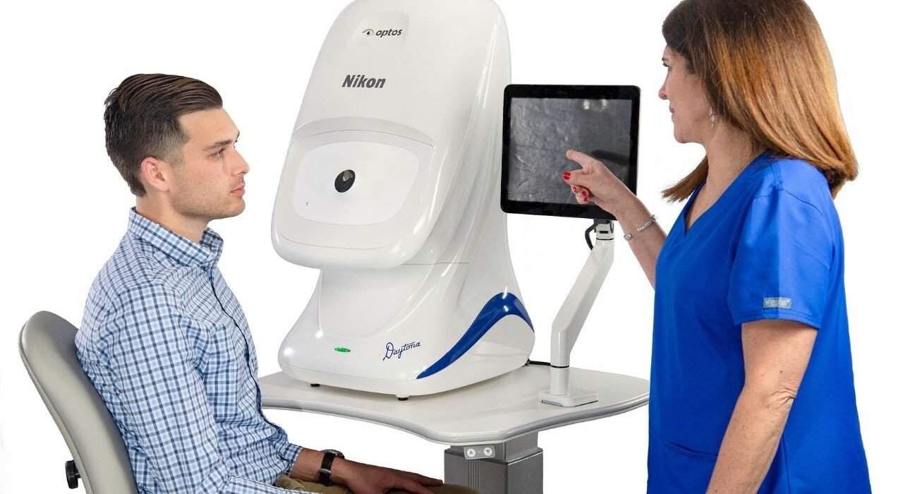
Unlocking 200° Pathology: A Clinical & Technical Guide to the Optos Daytona
In an era of rising global diabetic prevalence and an aging demographic, the burden on eye care facilities to rapidly and accurately diagnose posterior segment diseases has never been higher. Yet, for decades, the standard retinal camera has been constrained by fundamental optical limitations. Traditional digital fundus photography captures a narrow 30° to 45° field of view. While this is sufficient for documenting central maculopathies and optic nerve cupping, it leaves the vast majority of the peripheral retina completely unexamined in a single image.
Even when clinicians utilize the cumbersome, time-consuming ETDRS 7-field montage technique, they are only able to visualize approximately 75° to 90° of the retina. This central bias often leads to missed critical peripheral pathologies—such as peripheral diabetic ischemia, asymptomatic horseshoe tears, and early-stage melanomas—that frequently precede catastrophic central vision loss.
When evaluating a modern fundus camera for your hospital’s diagnostic lane, it is imperative to look beyond traditional flat-sensor imaging. The introduction of the Optos Daytona represents a paradigm shift in diagnostic ophthalmology. By moving clinics from basic posterior pole photography to true ultra widefield retinal imaging, the Daytona empowers retina specialists, comprehensive ophthalmologists, and progressive optometry centers to detect, document, and manage ocular diseases with unprecedented peripheral clarity.
This comprehensive B2B technical guide explores the optical engineering, clinical mechanics, workflow ROI, and advanced equipment comparisons necessary to successfully integrate the Optos Daytona into a high-throughput modern eye hospital.
The Optical Engineering Behind Optos Widefield Technology
The core physical limitation of a standard fundus camera is its reliance on white-light flash photography paired with flat optical sensors. A flat sensor cannot accurately map the spherical, concave interior of the ocular globe without introducing severe optical distortion and illumination drop-off at the extreme edges.
To overcome this geometric hurdle, Optos engineered a completely different imaging modality based on proprietary Scanning Laser Ophthalmoscopy (SLO) physics.
The Optos Daytona utilizes an advanced ellipsoidal mirror technology coupled with a virtual point source. Instead of projecting a broad, scattering flash of white light through the pupil, the system projects a sweeping, highly focused laser beam that technically pivots at the optical center of the eye. This virtual pivot point allows the scanning laser to sweep across a massive arc on the retinal surface without being clipped or blocked by the human iris.
Technical Performance Metrics:
200° Field of View (FOV): In a single, non-contact capture, the optos widefield system visualizes up to 82% of the total retinal surface area. This is a staggering geometric increase compared to the 15% captured by a standard 45° camera.
High-Speed Acquisition Engine: Diagnostic-quality high-resolution images are captured in less than 0.4 seconds. This micro-second speed significantly reduces the risk of motion artifacts, blink interference, and image blurring—common challenges when photographing elderly patients, children, or those with photophobia.
High-Resolution Output: The resulting optomap® image provides edge-to-edge optical clarity, allowing physicians to digitally zoom in on micro-pathologies (like isolated microaneurysms) in the far periphery without suffering from pixelation degradation.
Dual-Laser Scanning: Penetrating the Retinal Layers
While a traditional retinal camera captures a single, full-color superficial image, the Optos Daytona is fundamentally a tissue-penetrating, depth-layered diagnostic device. It utilizes a simultaneous dual-laser scanning engine that allows clinicians to virtually dissect retinal anatomy by depth, isolating specific layers for targeted pathological review.
Because different wavelengths of light penetrate ocular tissue to different depths, the optos ultra widefield system generates multiple distinct clinical views from a single 0.4-second scan:
532 nm Green Laser (Sensory Retina Focus): Green light is heavily absorbed by ocular melanin and hemoglobin. This laser scans from the superficial sensory retina down to the retinal pigment epithelium (RPE). It provides exceptionally high-contrast imaging of the retinal nerve fiber layer (RNFL), the macula, retinal vasculature, and superficial retinal hemorrhages. It is often referred to as a “red-free” equivalent, but with far higher contrast.
633 nm Red Laser (Choroidal Architecture): Red light penetrates much deeper into the tissue bed, bypassing the RPE to image the choroid directly. This specific wavelength is critical for visualizing deep vascular lesions, identifying choroidal nevi, monitoring choroidal melanomas, and evaluating the choroidal vasculature that underlies the retina.
Green Autofluorescence (FAF): The Daytona features a specialized Autofluorescence imaging mode. By stimulating lipofuscin—a metabolic waste product that accumulates in the RPE as photoreceptor cells degrade—the camera maps the underlying metabolic health of the retina. FAF is an indispensable tool for tracking geographic atrophy, monitoring the progression of dry Age-Related Macular Degeneration (AMD), and identifying early phenotypic signs of hereditary retinal dystrophies before they are clinically visible under standard light.
Eliminating the Dilation Bottleneck: The Non-Mydriatic ROI
From a hospital administration and operations perspective, the greatest hidden cost in a busy Outpatient Department (OPD) is patient wait time and clinical floor congestion. Traditional retinal evaluation requires pharmacological pupil dilation, forcing patients to sit in waiting bays for 30 to 45 minutes for mydriatic drops to take maximum effect. Furthermore, post-dilation blurred vision and severe photophobia significantly reduce the patient’s overall satisfaction and require them to arrange dedicated transportation.
The Optos Daytona is engineered as a true non mydriatic fundus camera, fundamentally redefining clinical throughput and delivering a massive Return on Investment (ROI) via time savings.
The Operational Advantages of Non-Mydriatic Imaging:
Imaging Through 2.0 mm Pupils: Because the virtual point source pivots the scanning lasers directly at the pupillary plane rather than trying to force a wide flash through it, the Daytona can effortlessly capture a full 200° optomap through un-dilated pupils as small as 2.0 mm. Routine pharmacological dilation is entirely bypassed for standard diagnostic screening.
Penetrating Media Opacities: Standard cameras rely on a bright white flash that scatters wildly when it hits a cataract, resulting in a washed-out, completely un-diagnostic image. The SLO laser technology of the Daytona cuts through moderate cataracts and vitreous opacities with minimal light scattering, delivering clear retinal maps where traditional photography fails.
Accelerated Patient Triage (The ROI Calculation): By eliminating the 40-minute dilation waiting period, an ophthalmic technician can seamlessly capture an optomap during the initial pre-examination workup alongside auto-refraction and tonometry. In a clinic seeing 50 patients a day, saving 30-40 minutes per patient clears waiting room bottlenecks instantly. The retina specialist has instant access to 200° of clinical data before the patient even enters the consultation room, allowing the doctor to increase daily patient volume by up to 25%.
Clinical Applications: Visualizing Peripheral Pathology
The clinical imperative for wide field retinal imaging is backed by decades of peer-reviewed data: sight-threatening diseases of the retina do not confine themselves to the central 45°. The Optos Daytona excels in identifying conditions that manifest silently in the periphery:
Diabetic Retinopathy (DR): Studies show that up to 30% of diabetic retinal lesions (microaneurysms, hemorrhages, and neovascularization) occur exclusively outside the standard ETDRS 7-field imaging area. The Daytona allows for accurate grading of peripheral ischemia and proliferative changes, directly influencing targeted panretinal photocoagulation (PRP) treatment plans and preventing unexpected vitreous hemorrhages.
Peripheral Retinal Tears & Detachments: Identifying asymptomatic horseshoe tears, retinal holes, or lattice degeneration in the far periphery is exceptionally difficult, even with a binocular indirect ophthalmoscope (BIO). The Daytona provides a comprehensive 200° map, ensuring these surgical emergencies are caught and sealed with a prophylactic laser before progressing to a full macula-off retinal detachment.
High Myopia Management: As global myopia rates soar, highly myopic eyes suffer from severe peripheral retinal thinning and stretching. The Daytona allows clinicians to accurately monitor peripheral retinal stretching, lattice degeneration, and pathological myopia progression over time.
Glaucoma Triage & Optic Nerve Evaluation: Beyond the periphery, the Daytona features robust central pole tools. By capturing modified dual-angle images, the software creates a 3D stereoscopic view of the optic nerve head, allowing glaucoma specialists to assess vertical cup-to-disc ratios and neuroretinal rim thinning.
Equipment Comparison: Optos Daytona vs. Optos California
For hospital procurement directors, choosing the correct optos fundus camera depends heavily on the specific sub-specialties offered at your facility. While the Daytona is a diagnostic powerhouse, Optos also offers a flagship surgical-grade model. How do you choose between the two?
The Optos California: The Surgical Retina Flagship
While the Daytona is the gold standard for high-volume general eye hospitals, comprehensive ophthalmology clinics, and premium optometry centers, the Optos California is specifically engineered for tertiary retina institutes, ocular oncology centers, and advanced surgical retina practices.
Key Differences to Consider:
Angiography Capabilities (FA & ICG): The most significant difference is that the Optos California offers Fluorescein Angiography (FA) and Indocyanine Green Angiography (ICG) in ultra-widefield. This allows vitreo-retinal surgeons to observe dynamic blood flow, vascular leakage, and choroidal neovascular membranes across the entire 200° periphery in real-time. The Daytona does not perform dye-based angiography (it offers Color, Red-Free, and Autofluorescence only).
Hardware & Optical Enhancements: The California features a redesigned optical head and modified hardware that makes capturing images of extreme peripheral pathology (e.g., pars plana tumors) slightly easier in highly uncooperative or complex surgical patients.
Target Deployment:
Choose the Daytona if your goal is rapid OPD screening, diabetic retinopathy management, general pathology documentation, and maximizing non-mydriatic patient throughput.
Choose the California if you operate a dedicated vitreo-retinal surgical center requiring deep vascular angiography and ocular tumor management.
Software Integration: OptosAdvance™ & Enterprise Connectivity
Advanced hardware is only as effective as the software managing its data. The Optos Daytona integrates directly with OptosAdvance™, a comprehensive, browser-based image review and management software designed for enterprise clinical environments.
Dynamic Image Manipulation: Clinicians can easily toggle between the composite color image, the isolated green laser view, the red laser view, and autofluorescence with a single click.
Digital Auto-Montage (220° View): While a single capture provides 200°, the software can automatically stitch together superior, inferior, nasal, and temporal steered images to create an unprecedented 220° view of the retina, covering nearly the entirety of the ocular interior.
Longitudinal Tracking: The software allows for precise, side-by-side historical comparison of optomap images. This is vital for monitoring geographic atrophy expansion or verifying that a peripheral diabetic hemorrhage is resolving post-injection.
EHR and HIS Integration: The Daytona is fully DICOM compatible. Images and diagnostic reports can be pushed instantly to local Hospital Information Systems (HIS), Electronic Health Records (EHR), or cloud-based PACS servers, enabling seamless multi-location teleophthalmology consultations and remote grading.
Why Source the Optos Daytona from Mediscience Devices?
Procuring an advanced optos widefield system is a major capital expenditure that requires partnering with a highly certified biomedical distribution network. Purchasing high-tier diagnostic equipment from unauthorized vendors or grey-market importers introduces severe institutional risks, including uncalibrated optical engines, lack of software license transfers, and absent warranty support.
As Eastern and North-Eastern India’s premier authorized distributor of ophthalmic diagnostic equipment for over 27 years, Mediscience Devices ensures your capital investment is fully protected.
The Mediscience B2B Guarantee:
Turnkey Installation: Our factory-trained biomedical engineers manage the complete physical deployment, network IP configuration, DICOM integration, and exact optical calibration of the Daytona unit within your hospital’s infrastructure.
Clinical Application Training: Technology is useless without proper operation. We provide comprehensive on-site training for your optometrists and clinical technicians, ensuring your staff knows exactly how to position patients, navigate the software, and maximize the image capture rate.
Uninterrupted Service Level Agreements (SLAs): We maintain a local depot of spare components and provide rapid on-site technical support across Kolkata, West Bengal, and the broader Eastern Indian region, guaranteeing near-zero clinical downtime.
Conclusion: Upgrade to the Modern Standard of Retinal Care
The Optos Daytona is not merely a high-end camera; it is a fundamental transformation of the diagnostic workflow. By seamlessly combining 200° ultra widefield retinal imaging, multi-laser depth penetration, and a rapid non-mydriatic screening process, the Daytona enables clinicians to uncover sight-threatening peripheral pathology that traditional cameras leave completely in the dark.
Whether diagnosing early-stage diabetic retinopathy or monitoring peripheral lattice degeneration, equipping your OPD with the Daytona drastically accelerates patient throughput while simultaneously elevating the standard of clinical care you provide.
Elevate your hospital’s diagnostic capabilities and protect your patients from undetected peripheral retinal diseases.
Contact the B2B Institutional Sales Division at Mediscience Devices today to request an official quotation, explore comprehensive Annual Maintenance Contracts (AMC), and schedule an exclusive on-site demonstration of the Optos Daytona.
People Also Ask
Yes. Unlike a traditional retinal camera that requires a large, dilated pupil to allow a bright flash to pass through, the Daytona utilizes targeted Scanning Laser Ophthalmoscopy (SLO). This technology allows the device to capture a comprehensive 200° image through an un-dilated pupil as small as 2.0 mm. This true non mydriatic fundus camera capability eliminates the 40-minute wait time for dilating drops during routine diagnostic screenings.
A conventional digital fundus camera typically captures only a 30° to 45° field of view, visualizing roughly 15% of the retina (mostly the macula and optic nerve). In contrast, the Optos Daytona delivers ultra widefield retinal imaging, capturing a 200° panoramic view that covers up to 82% of the retina in a single 0.4-second scan. This allows clinicians to easily detect peripheral pathologies like diabetic ischemic changes and asymptomatic retinal tears.
Yes. Traditional white-light photography often scatters when hitting a cataract or vitreous haze, resulting in a washed-out, undiagnostic image. The narrow-band dual-laser technology used in the optos fundus camera is highly effective at cutting through moderate cataracts and media opacities with minimal light scattering, providing clear, diagnostic-quality images where standard imaging modalities fail.
The Daytona is fully integrated with OptosAdvance™ image management software, which is 100% DICOM compliant. This ensures that every piece of wide field retinal imaging data can be seamlessly pushed to your hospital's Picture Archiving and Communication System (PACS), Hospital Information System (HIS), or Electronic Health Records (EHR) for instant multi-department review and teleophthalmology consultations.
Healthcare institutions can procure certified, factory-warranted eye clinic equipments directly through authorized regional distributors like Mediscience Devices in Kolkata. Working with an established distributor ensures authentic pricing, professional installation, staff training, and rapid local technical support across Eastern and North-Eastern India.