Digital Fundus Camera in Nepal: A Complete Buyer’s Guide for Hospitals & Eye Clinics
Eye care delivery in Nepal is advancing rapidly. Across tertiary eye hospitals, medical colleges, private eye centers, and community screening networks, clinicians face a rising burden of preventable blindness driven by diabetic retinopathy, glaucoma, and age-related macular degeneration (AMD). Because early-stage posterior segment diseases often progress silently without noticeable vision loss, routine photographic documentation of the retina and optic nerve head has become essential for modern ophthalmology and optometry practices.
At the center of posterior segment screening sits the digital fundus camera. Unlike traditional direct or indirect ophthalmoscopy, which relies solely on fleeting real-time observation, modern fundus photography equipment captures high-resolution digital images of the retina, macula, optic disc, and retinal vasculature. These visual records allow eye care professionals to track subtle disease progression over time, educate patients directly at the chairside, and share images remotely with retinal specialists for tele-ophthalmology consultations.
For hospital directors, clinic owners, and medical equipment distributors evaluating a digital fundus camera for deployment in Nepal, choosing the right system requires balancing optical resolution, non-mydriatic pupil capabilities, portability, and long-term technical support.
This comprehensive buyer’s guide explores the optical engineering behind modern retinal imaging, provides in-depth technical profiles of leading models in Mediscience Devices’ fundus camera category, analyzes commercial investment factors, and outlines how Mediscience supplies healthcare providers across Nepal.
Understanding Digital Fundus Camera Technology: Non-Mydriatic vs. Mydriatic Imaging
A retinal camera (fundus camera) is a specialized low-power microscope attached to an optical camera system designed to photograph the interior surface of the eye through the pupil. When evaluating systems, clinical procurement teams will encounter two primary operational architectures:
Why Non-Mydriatic Imaging Transforms OPD Throughput:
Zero Dilation Bottlenecks: Traditional fundus photography required instilling mydriatic drops and waiting 30 to 40 minutes for pupil dilation, creating waiting room congestion and causing temporary photophobia and blurred near vision for patients. A non-mydriatic digital fundus camera uses harmless infrared illumination to align and focus on the retina in dim ambient light, triggering a brief white LED flash only at the exact millisecond of image capture.
Small-Pupil Screening Capability: Elderly patients, diabetic individuals, and patients with physiological miosis frequently present with constricted pupils. Advanced non-mydriatic systems capture crisp, reflection-free retinal images through pupils as small as 2.5 mm to 3.3 mm.
Tele-Ophthalmology & AI Screening Integration: Digital retinal images captured by technicians in primary clinics or mobile outreach camps can be transmitted wirelessly to retinal specialists or processed through Artificial Intelligence (AI) screening algorithms to flag referable diabetic retinopathy immediately.
Detailed Product Profiles: Featured Fundus Cameras from Mediscience
Selecting the ideal fundus camera requires matching your clinic’s patient demographic, spatial footprint, and mobility needs with the right hardware platform. Mediscience Devices supplies a specialized range of handheld and compact tabletop retinal imaging systems tailored for clinical practices and outreach programs in Nepal.
1. Mocular Model CFC-X Digital Portable Fundus Camera
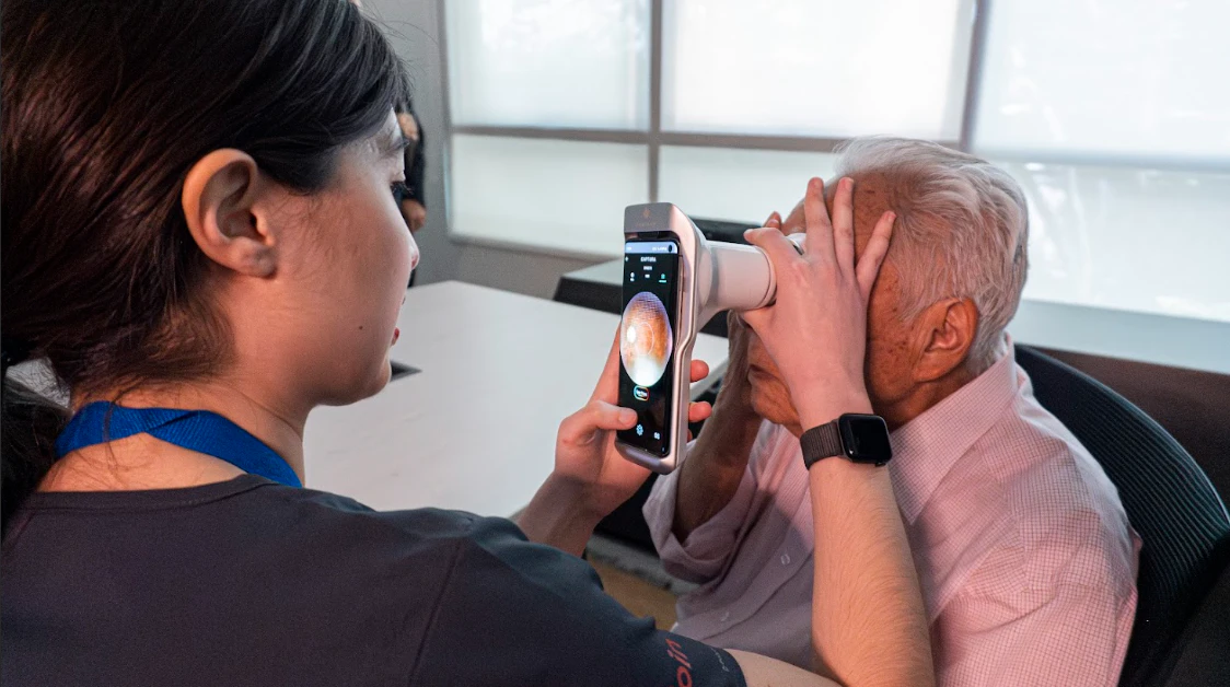
The Mocular Model CFC-X Digital Portable Fundus Camera is a truly wireless, user-friendly handheld non-mydriatic portable fundus camera engineered to bring high-definition retinal photography beyond fixed examination rooms. Weighing less than 600 grams, the CFC-X is built for exceptional mobility across busy outpatient clinics, pediatric bays, bedside hospital ward rounds, and community eye screening camps. Equipped with a high-resolution 12-megapixel capture sensor, white LED and infrared LED illumination, and a 24 mm working distance, it captures crisp 50° field-of-view retinal photographs through un-dilated pupils as small as 2.5 mm. Furthermore, its multi-point internal fixation system uses artificial star-point guidance technology to auto-focus across multiple angles, covering an expanded field of view up to 90°.
Designed as a self-contained mobile workstation, the Mocular CFC-X features a 4.7-inch anti-glare multi-touch display powered by a 4-core 2.2 GHz processor with 4 GB RAM and 64 GB internal storage. Clinicians can adjust focus manually or rely on rapid autofocus, review images chairside with patients, and utilize built-in AI diagnostic report support to help identify retinal anomalies. Connectivity is seamless with integrated Wi-Fi, Bluetooth, USB 3.0, Type-C, SD card slot, 4G, and NFC support, alongside DICOM compatibility for direct integration into hospital management networks and wireless printing. With a rapid charging time of under 2 hours and an extended battery operation time exceeding 5 to 8 hours per charge, the Mocular CFC-X arrives in a durable carrying case ready for continuous field or clinic deployment.
2. Volk Fundus Camera
The Volk Fundus Camera combines legendary Volk optical glass engineering with modern portable retinal imaging technology, delivering reflection-free, high-contrast fundus photographs across fixed clinics, mobile health units, and outreach programs. Built around a high-resolution 3-CCD (Charge-Coupled Detector) capture sensor with 10-line-pair optical resolution and a 40 mm working distance, this portable retinal camera captures sharp color and red-free retinal images with true-to-life color fidelity. The optical engine features a wide diopter compensation focus range from -20D to +20D, allowing operators to achieve crisp focus effortlessly across high myopes, hyperopes, and post-surgical patients using either automatic or manual focusing controls.
Engineered for clinical versatility, the Volk Fundus Camera supports a standard 5 mm pupil diameter as well as a dedicated 4.3 mm small-pupil mode, paired with four external fixation targets and automatic internal fixation operation to guide patient gaze accurately. Its low-intensity LED light source features 20 to 25 adjustable flash levels to maximize patient comfort while preserving delicate retinal detail. Operators benefit from onboard Image Quality Analysis (IQA) to verify image clarity immediately upon capture, a 15-inch LED display interface, and versatile mounting flexibility that allows the camera to be used handheld or mounted directly onto a slit lamp. With integrated Wi-Fi, Bluetooth, and USB-C data transfer capabilities, the Volk Fundus Camera generates instant, shareable patient reports for seamless referral workflows and clinical documentation.
3. 3 Nethra Fundus Camera
The 3 Nethra Fundus Camera (3nethra Classic) is a compact, multifunctional non-mydriatic digital fundus camera designed to capture evenly illuminated, undistorted images of both the anterior and posterior segments of the human eye. Featuring a high-resolution 6.4-megapixel camera sensor, a 45° field of view, and an optical resolution of 8 to 14 microns, the 3 Nethra delivers detailed digital documentation through pupils as small as 3.3 mm without requiring routine pharmacological dilation. Its compact structural footprint (520 mm H × 420 mm L × 400 mm W) and modular weight configuration—comprising a 3.4 kg tricam optical head and a 7.7 kg stand for a total weight of 11.1 kg—make it easy to integrate into busy outpatient consultation lanes or transport for community screening initiatives.
A standout advantage of the 3 Nethra Fundus Camera is its true multifunctional diagnostic capability within a single workstation. Beyond standard non-mydriatic posterior retinal photography and peripheral retinal imaging, the system captures high-clarity anterior segment photographs and performs specialized dry-eye imaging including meibography. Operating on ultra-low 5 to 10 W DC power consumption, the system delivers fast examination times with an intuitive user interface that requires minimal technician training. Certified with US-FDA clearance, CE marking, and Health Canada registration, the 3 Nethra is fully compatible with multiple AI-assisted retinal screening platforms and telemedicine networks, making it a dependable workhorse for eye hospitals, diabetic screening centers, and tele-ophthalmology programs.
Master Technical Comparison Matrix
To assist hospital procurement committees, optometrists, and clinical directors in selecting the right retinal imaging system, the table below compares the core technical parameters of these three platforms:
| Technical Parameter | Mocular Model CFC-X | Volk Fundus Camera | 3 Nethra Fundus Camera |
| Form Factor | Wireless Handheld Portable (< 600 g) | Portable Handheld / Slit-Lamp Mountable | Compact Tabletop Workstation (11.1 kg total) |
| Imaging Mode | Non-Mydriatic | Mydriatic (with 4.3 mm Small-Pupil Mode) | Non-Mydriatic & Multifunctional |
| Sensor Resolution | 12 Megapixels | High-Resolution 3-CCD Sensor | 6.4 Megapixels (8–14 microns resolution) |
| Field of View (FOV) | 50° (Up to 90° with multi-point fixation) | Wide-Field Volk Optics | 45° |
| Minimum Pupil Size | 2.5 mm | 4.3 mm (Small-Pupil Mode) / 5.0 mm (Normal) | 3.3 mm |
| Focus & Compensation | Manual & Auto Focus | Manual & Auto Focus (-20D to +20D) | Precision Optical Alignment |
| Specialized Capabilities | Built-in AI report support, 4G, NFC, DICOM | Red-free imaging, Onboard IQA, 20–25 flash levels | Posterior, Anterior & Dry-Eye Meibography |
| Connectivity | Wi-Fi, Bluetooth, USB 3.0, Type-C, SD, 4G | Wi-Fi, Bluetooth, USB-C | AI Systems & Telemedicine Platforms |
Understanding Fundus Camera Price & Total Cost of Ownership (TCO)
When evaluating a fundus camera price proposal for an eye clinic or hospital department, purchasing managers must look beyond the upfront hardware invoice to analyze the Total Cost of Ownership (TCO) and return on clinical investment.
Commercial Value Drivers Behind Equipment Selection:
Non-Mydriatic Workflow Efficiency: Eliminating routine dilation drops allows clinics to screen more patients per hour, reduces consumable eye-drop expenses, and improves patient satisfaction.
Multifunctional Diagnostic Yield: Selecting a platform like the 3 Nethra Fundus Camera—which combines posterior retinal imaging with anterior photography and dry-eye meibography—maximizes capital utilization by fulfilling multiple diagnostic needs from one footprint.
Outreach & Tele-Ophthalmology Scalability: Investing in a lightweight portable fundus camera like the Mocular Model CFC-X allows healthcare organizations to run mobile diabetic screening camps and rural outreach programs without transporting heavy tabletop tables.
Software & AI Compatibility: Open connectivity (Wi-Fi, USB, DICOM, and AI screening integration) prevents expensive proprietary software lock-ins and ensures seamless record archiving.
Reliable Supply & Technical Support for Nepal
Sourcing specialized fundus photography equipment requires working with a trusted supplier that ensures genuine hardware, safe transit, and dependable technical assistance. Mediscience Devices supplies ophthalmic hospitals, private eye clinics, diagnostic centers, and optical practices across Nepal with direct access to top-tier retinal imaging systems.
Why Eye Care Providers in Nepal Partner with Mediscience
Authentic Global Brands: Direct supply of genuine Mocular, Volk, and 3 Nethra imaging platforms backed by manufacturer warranties.
Complete Supply Management to Nepal: We coordinate seamless cross-border shipping, customs documentation, and secure protective packaging to deliver delicate optical equipment safely to your facility in Nepal.
Clinical & Technical Guidance: Our biomedical specialists assist your clinical team with device setup, image capture workflows, wireless printing configuration, and tele-ophthalmology integration.
Turnkey Ophthalmic Bundling: Hospitals and clinics can bundle their digital fundus camera purchase alongside slit lamps, auto refractometers, tonometers, ultrasound A/B-scans, and surgical microscopes under a single streamlined procurement order.
Conclusion
Integrating a high-precision digital fundus camera into your clinical practice elevates patient care by enabling early detection of retinal pathologies, streamlining patient education, and supporting modern tele-ophthalmology workflows. Whether your facility requires the ultra-lightweight wireless mobility of the Mocular Model CFC-X Digital Portable Fundus Camera, the optical precision of the Volk Fundus Camera, or the multifunctional non-mydriatic versatility of the 3 Nethra Fundus Camera, Mediscience Devices supplies authentic retinal imaging solutions across Nepal.
Contact the Sales Division at Mediscience Devices today to request an official quotation, discuss institutional pricing for Nepal, and schedule a product consultation for your clinic.
People Also Ask
A non-mydriatic digital fundus camera captures detailed photographs of the retina without requiring pharmacological eye drops to dilate the pupil. This eliminates the 30-to-40-minute waiting period for dilation, improves patient comfort, and significantly increases daily clinic throughput.
Yes. Handheld models like the Mocular Model CFC-X weigh under 600 grams, operate on rechargeable batteries for over 5 hours, and include wireless Wi-Fi and 4G connectivity—making them ideal for mobile outreach programs, school screenings, and remote health posts.
Beyond standard 45° non-mydriatic posterior retinal photography, the 3 Nethra Fundus Camera supports anterior segment imaging and dry-eye imaging (meibography), giving clinics multiple diagnostic capabilities in one compact device.
Yes. In addition to handheld portable use, the Volk Fundus Camera features slit-lamp mounting compatibility, allowing clinicians to stabilize the camera on an existing slit lamp stand during routine OPD examinations.