Optometry Clinic Setup: Complete Equipment, Diagnostic Lane & Procurement Guide
Establishing a modern practice or upgrading an existing facility requires balancing clinical precision, patient throughput, space utilization, and financial planning. Whether you are expanding a retail optical practice into a full-scope optometry clinic, establishing a multi-specialty advanced eye clinic, or outfitting an integrated ophthalmology clinic setup, selecting the right diagnostic infrastructure sets the foundation for your practice’s success.
In contemporary optometry, patients expect comprehensive vision care that goes beyond simple subjective refraction. Modern diagnostic lanes must deliver rapid screening for refractive errors, early glaucoma detection, corneal pathology evaluation, and posterior segment retinal health mapping.
For clinical entrepreneurs, hospital directors, and eye care professionals evaluating the equipment required for an eye clinic, this comprehensive guide details essential diagnostic machinery, structures efficient lane layouts, analyzes the eye clinic setup cost, and provides a strategic procurement roadmap.
Planning Your Optometry Clinic Setup: Spatial & Workflow Architecture
Before procuring optical equipment, clinic directors should map out the operational layout to ensure seamless patient traffic flow. A well-designed optometry clinic typically divides its floor space into distinct functional zones:
Pre-Testing Screening Bay: Outfitted with automated, high-speed instruments (such as autorefractors and non-contact tonometers) that technicians operate before the patient enters the main consultation room.
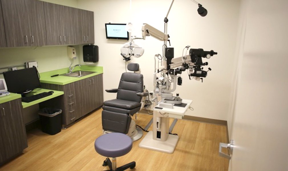
Primary Examination Lane: The core diagnostic hub where the practitioner performs detailed slit-lamp biomicroscopy, subjective refraction, and fundus evaluations.
Specialized Diagnostic Suite: Reserved for advanced testing such as fundus photography, optical coherence tomography (OCT), perimeter visual fields, and A/B-scan ultrasonography.
Core Diagnostic Equipment Required for an Eye Clinic
Building an efficient practice requires choosing high-precision instruments that deliver consistent, repeatable readings. Below are the essential diagnostic workstations that anchor every professional diagnostic lane:
1. Slit Lamp Biomicroscope: Anterior Segment Diagnostic Anchor
A high-resolution Slit Lamp is the single most critical biomicroscope in any diagnostic bay. Combining an intense illumination light engine with a Galilean optical system, it allows clinicians to examine cross-sectional optical slices of the cornea, anterior chamber, iris, crystalline lens, and anterior vitreous.
Selection Criteria: Choose between 3-step or 5-step Galilean drum magnification systems with Apochromatic glass optics. Ensure the frame supports applanation tonometer mounts, diffuse background lighting, and digital camera beam-splitters for patient record archiving.
2. Auto Refractometer / Keratometer: Automated Refractive Gatekeeper
An automated Auto Refractometer serves as the initial diagnostic workhorse. By projecting near-infrared light through the pupil onto the retina, it measures sphere, cylinder, axis, and corneal curvature radii ($K_1, K_2$) in seconds.
Selection Criteria: Look for systems equipped with Hartmann-Shack wavefront aberrometry, dual-CCD camera sensors, auto-fogging scenic targets to prevent accommodation spasms, and small-pupil measurement capabilities (down to 2.0 mm).
3. Tonometer: Intraocular Pressure (IOP) & Glaucoma Screening
Glaucoma screening requires precise measurement of intraocular fluid pressure. Integrating a reliable Tonometer is essential for detecting ocular hypertension before irreversible optic nerve damage occurs.
Selection Criteria: Practices can select from desktop non-contact (air-puff) tonometers for high-volume screening bays, handheld rebound tonometers (requiring no anesthesia), transpalpebral devices, or classic applanation units. For a detailed breakdown of pressure testing mechanics, explore specialized tonometer eye pressure platforms.
4. Ophthalmoscope & Fundus Camera Setup: Posterior Segment Mapping
Evaluating the retina, optic nerve head, macula, and retinal vasculature requires high-performance fundus imaging tools. Outfitting your practice with an advanced Fundus Camera setup allows clinicians to screen for diabetic retinopathy, macular degeneration, and hypertensive changes.
Selection Criteria: Non-mydriatic digital fundus camera systems allow rapid high-definition retinal photography through un-dilated pupils, streamlining patient throughput and supporting tele-ophthalmology consultations.
5. Trial Lens Set & Trial Frame: Subjective Refraction Baseline
While automated refractors provide accurate baseline data, final optical prescriptions require subjective verification using a precision Trial Lens Set paired with an ergonomic Trial Frame.
Selection Criteria: Select reduced-aperture, anti-reflective coated glass trial lenses housed in durable aluminum or velvet-lined carrying cases. Pair them with lightweight, fully adjustable trial frames featuring smooth interpupillary distance (PD), axis, cell rotation, and vertex distance adjustments.
Advanced Eye Clinic Expansion: Specialized Equipment
To elevate your practice into an advanced eye clinic or full-scope surgical triage center, consider expanding your diagnostic suite beyond basic refraction and anterior screening:
Digital Auto Lensmeter: Automatically verifies spectacle lens powers, progressive addition heights (PALs), prism deviations, and UV/Blue-light coating transmission percentages within seconds.
Ophthalmic Ultrasound (A-Scan / B-Scan): Essential for calculating Intraocular Lens (IOL) power prior to cataract surgery and imaging the posterior segment through dense media opacities (such as mature cataracts or vitreous hemorrhage).
Automated Visual Field Perimeter: Maps peripheral retinal sensitivity to diagnose, stage, and monitor open-angle and angle-closure glaucoma progression.
For a deeper look into prioritizing your clinical purchases, read our guide on 5 Must-Have Eye Clinic Equipments.
Understanding the Eye Clinic Setup Cost
Evaluating the overall eye clinic setup cost requires breaking down capital expenditure (CapEx) into distinct equipment tiers. Understanding these tiers helps clinical entrepreneurs align hardware selection with available budgets and projected patient volumes:
Commercial Equipment Tiers
Primary OPD Setup (Entry-to-Mid Tier): Includes a manual 2-step or 3-step slit lamp, standard tabletop autorefractometer, trial lens set with frame, chart projector/LCD chart, and a manual or rebound tonometer. This tier provides a budget-conscious entry point for new independent practices.
Comprehensive Diagnostic Setup (Professional Tier): Features a 5-step Apochromatic slit lamp with applanation tonometer, dual-mode auto ref-keratometer, digital lensmeter, non-contact air-puff tonometer, and digital vision chart. Ideal for busy urban eye centers and optical chains.
Advanced Tertiary Clinic Setup (Flagship Tier): Incorporates top-tier Japanese or German Apochromatic slit lamps, non-mydriatic fundus cameras, A/B ultrasound scanners, digital auto phoropters, and computerized perimeters. Designed for multi-specialty centers and surgical referral hubs.
Master Product Comparison Matrix for Clinic Setup
To assist procurement committees, hospital boards, and practice owners in evaluating essential diagnostic instruments, the table below compares key operational and clinical parameters:
| Equipment Category | Primary Clinical Function | Key Features to Inspect | Ideal Zonal Placement | Relative CapEx Tier |
| Slit Lamp Biomicroscope | Anterior segment micro-inspection | 3/5-Step Galilean drum, LED lighting, tonometer mount | Main Consultation Lane | Core Essential |
| Auto Refractometer | Automated objective refraction & keratometry | Wavefront sensor, 2.0mm pupil threshold, auto-fog target | Pre-Testing Screening Bay | Core Essential |
| Tonometer | Intraocular Pressure (IOP) screening | Non-contact air puff, rebound, transpalpebral, or applanation | Pre-Testing / Main Lane | Core Essential |
| Fundus Camera Setup | Retinal & optic nerve imaging | Non-mydriatic capture, HD resolution, DICOM integration | Advanced Diagnostic Suite | Advanced Upgrade |
| Trial Lens Set & Frame | Subjective trial lens refinement | Reduced aperture, anti-reflective glass, lightweight frame | Main Consultation Lane | Core Essential |
Sourcing & Procurement: Direct Local & Regional Supply via Mediscience
Procuring diagnostic equipment requires choosing a distribution partner that combines technical expertise, transparent pricing, and dependable after-sales technical support.
Why Healthcare Facilities Partner with Mediscience Devices:
Complete Turnkey Setup: Mediscience supplies the entire spectrum of diagnostic and surgical tools—allowing buyers to consolidate full-clinic orders under a single invoice.
Factory-Trained Technical Engineering: Certified biomedical engineers handle physical assembly, optical alignment, network integration, and staff operational training on site.
Hassle-Free Regional Logistics: With dedicated local operations in Nepal and established export routes to the UAE and neighboring markets, Mediscience manages cross-border customs clearance, local billing, and doorstep fulfillment.
Conclusion
Planning a successful optometry clinic setup is an investment in clinical diagnostic accuracy, workflow speed, and long-term practice growth. By combining essential baseline tools—such as high-resolution slit lamps, wavefront auto refractometers, precision tonometers, fundus imaging suites, and trial optics—you create an environment that delivers exceptional eye care.
Whether you are opening a new optical consultation lane in India or sourcing turnkey clinic equipment across Nepal, the UAE, or international markets, Mediscience Devices provides authentic global technology, competitive pricing, and technical support.
Contact the Clinical Procurement Division at Mediscience Devices today to request a customized equipment quote, discuss turnkey clinic packages, and schedule a consultation with our biomedical specialists.
People Also Ask
A standard primary consultation lane requires a room measuring approximately $8\text{ ft} \times 10\text{ ft}$ (or a 3-meter optical length) to comfortably accommodate a patient chair, motorized refraction stand, slit lamp, visual chart distance, and practitioner workspace.
If your clinic expects high daily patient volume in a pre-testing bay, a desktop non-contact (air-puff) tonometer offers rapid, non-invasive screening without drops. If you plan to conduct pediatric exams, home visits, or mobile screening camps, a portable rebound tonometer provides high portability without requiring anesthesia.
Yes. Many 3-step and 5-step Galilean drum slit lamps feature modular optical heads that accept camera beam splitters, HD camera adapters, and background illuminators, allowing you to upgrade to a digital archiving suite as your clinic grows.
A standard autorefractometer measures objective refractive error (sphere, cylinder, axis). An auto ref-keratometer combines two tests in one, measuring refractive error alongside corneal curvature radii ($K_1, K_2$), which is essential for contact lens fittings and pre-op cataract screening.