The Ultimate Buyer’s Guide to Corneal Topographer in India
In modern ophthalmology and refractive surgery, diagnostic accuracy at the anterior segment is the foundational pillar of clinical success. While a standard auto refractometer remains an essential screening tool for measuring baseline objective sphere and cylinder values, it captures data from only the central 3 mm of the cornea. For clinicians diagnosing irregular astigmatism, screening for ectatic corneal disorders like subclinical keratoconus, fitting specialty contact lenses, or planning premium Toric Intraocular Lenses (IOLs), basic keratometry falls short.
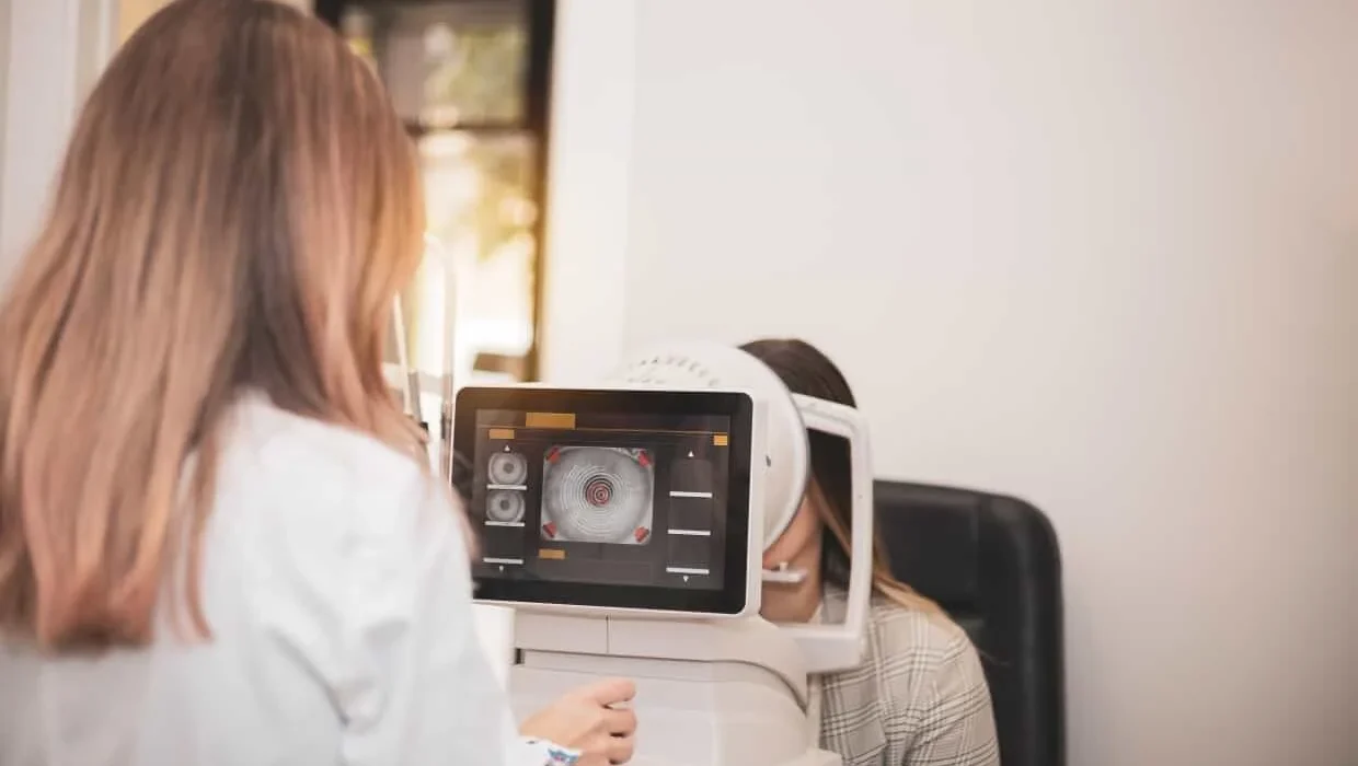
Investing in an advanced corneal topographer expands diagnostic vision across the entire corneal surface. By projecting thousands of data points onto the anterior surface, a modern corneal topography machine creates high-resolution axial, tangential, and elevation maps that expose subtle structural abnormalities long before they become visible under a slit lamp.
For hospital directors, Chief Medical Officers (CMOs), and clinic founders evaluating capital medical devices, selecting the best corneal topographer requires balancing optical precision, multi-modal versatility, spatial efficiency, and Total Cost of Ownership (TCO).
As one of Eastern and North-Eastern India’s most trusted equipment suppliers for over 27 years, Mediscience Devices provides this technical procurement guide to help healthcare facilities choose the right anterior segment imaging systems.
The Clinical Mandate: Why Every Modern Eye Hospital Needs a Corneal Topography Machine
The evolution of cataract and refractive surgery has transformed patient expectations. Cataract surgery is no longer viewed merely as a restorative procedure, but as a refractive one. Achieving emmetropia requires precise mapping of corneal shape, power, and tear film stability.
Essential Clinical Applications:
Pre-Operative Refractive Surgery Screening: Ruling out subclinical keratoconus, pellucid marginal degeneration (PMD), and irregular astigmatism prior to LASIK, PRK, or SMILE procedures.
Premium Cataract Surgery & Toric IOL Planning: Accurately measuring the steep and flat corneal axes and corneal astigmatism to ensure precise alignment of Toric IOLs.
Specialty Contact Lens Fitting: Generating custom sagittal height and elevation maps required to fit Rigid Gas Permeable (RGP), Ortho-K, and scleral lenses.
Ocular Surface & Tear Film Analysis: Evaluating tear film break-up times and corneal epithelial integrity, ensuring dry eye disease is treated prior to surgical biometry.
Core Diagnostic Modalities in Anterior Segment Analysis
When evaluating a corneal topography device, procurement committees must understand the primary optical sensing engines available in the market.
Placido Disc Systems: Projects concentric rings of light onto the tear film. The reflected ring pattern is analyzed to measure curvature across thousands of points. Placido systems offer exceptionally high resolution for anterior corneal curvature and tear film stability analysis.
Shack-Hartmann Wavefront Aberrometry: Maps optical ray deviations as light travels through the cornea and crystalline lens, quantifying lower-order (sphere, cylinder) and higher-order aberrations (coma, trefoil, spherical aberration).
Multi-Modal Diagnostic Integration: Modern practices increasingly prefer multi-functional units that combine auto-refraction, keratometry, topography, and ocular surface analysis into a single physical footprint.
Featured Diagnostic Systems: Interlinking Key Category Models
To streamline facility planning, Mediscience Devices highlights two flagship diagnostic systems representing peak engineering in multi-modal topography and specialized ocular surface analysis.
Rexxam AR-Kerato Topographer REF-TOPO RET-700
For high-volume Outpatient Departments (OPDs) seeking maximum spatial efficiency and rapid patient throughput, the Rexxam AR-Kerato Topographer REF-TOPO RET-700 is a premier 3-in-1 workstation.
3-in-1 Workstation Integration: Combines a high-precision auto refractometer, an auto keratometer, and a Placido disc corneal topographer into a single device. Patients complete objective refraction, curvature readings, and 3D corneal mapping in a single seating.
Japanese Optical Precision: Manufactured by Rexxam (formerly Shin-Nippon), it utilizes multi-coated optical arrays and advanced auto-fogging targets to relax patient accommodation, capturing accurate refractive readings even through small pupil diameters (down to 2.0 mm).
Topographic Mapping Suite: Generates detailed axial, tangential, and refractive maps, enabling instant screening for irregular astigmatism, corneal dystrophies, and contact lens fitting parameters.
Retro-Illumination Capabilities: Allows clinicians to capture high-contrast images of crystalline lens opacities, providing objective visual documentation for cataract grading and patient counseling.
MediWorks Dry Eye Analyzer DEA-520
In modern anterior segment care, corneal topography and dry eye diagnostics are deeply interconnected. An unstable tear film distorts Placido ring reflections, leading to inaccurate topographic maps and false IOL power calculations. The MediWorks Dry Eye Analyzer DEA-520 is a specialized, high-definition ocular surface topographer engineered to deliver comprehensive dry eye evaluations.
Why the MediWorks DEA-520 is Essential for Your Topography Suite:
The MediWorks Dry Eye Analyzer DEA-520 elevates ocular surface care by transforming qualitative dry eye symptoms into quantitative, reproducible data:
Non-Invasive Tear Film Break-Up Time (NIBUT): Utilizing grid projection algorithms, the DEA-520 measures first and average break-up times automatically without fluorescein dye instillation, preserving natural tear film dynamics.
Infrared Meibography & Gland Loss Analysis: Features specialized infrared light sensors to image the Meibomian gland architecture in the upper and lower eyelids. The integrated software automatically calculates the percentage of gland loss, allowing clinicians to stage Meibomian Gland Dysfunction (MGD).
Lipid Layer & Tear Meniscus Height Measurement: Evaluates lipid layer thickness via optical interference patterns and measures Tear Meniscus Height (TMH) down to the millimeter, helping differentiate evaporative dry eye from aqueous-deficient dry eye.
Ocular Redness & Corneo-Conjunctival Staining: Automatically quantifies bulbar and ciliary conjunctival hyperemia using AI-assisted image recognition, providing objective baselines to monitor therapeutic progress.
Pre-Surgical Ocular Surface Optimisation: Deploying the DEA-520 alongside your corneal topography machine ensures that dry eye disease is diagnosed and treated before refractive or cataract surgery, guaranteeing pristine corneal reflections for surgical planning.
Enterprise Comparison Matrix
To assist hospital purchasing committees and lead surgeons in selecting the best corneal topographer configuration for their facility, the matrix below compares primary operational parameters:
| Feature / Specification | Rexxam REF-TOPO RET-700 | MediWorks DEA-520 | Standalone Tabletop Topographer |
| Primary Clinical Focus | Refraction, Keratometry & Corneal Topography | Comprehensive Ocular Surface & Dry Eye Analysis | Dedicated Corneal Topography & Ectasia Mapping |
| Diagnostic Engine | 3-in-1 Multi-Modal Workstation | High-Definition Optical Surface Analyzer | Placido Disc / Scheimpflug Camera |
| Auto-Refraction Included? | Yes (Built-in Auto Refractometer) | No (Focuses on Ocular Surface) | No |
| Specialized Meibography? | Basic Anterior Imaging | Yes (Infrared Gland Loss Mapping) | Optional Add-on |
| NIBUT Measurement? | Standard Tear Stability Check | Yes (Automated Non-Invasive Break-Up) | Basic Tear Break-Up |
| OPD Floor Footprint | Extremely Compact (1 Tabletop Unit) | Dedicated Diagnostic Station | Single Tabletop Unit |
| Ideal Clinical Placement | High-Volume OPD Lanes, General Eye Clinics | Premium Dry Eye Clinics, Refractive Suites | Refractive Surgery Bays, Corneal Specialty |
B2B Procurement Checklist for Hospital Purchasing Directors
Selecting capital medical devices requires rigorous evaluation past basic equipment specifications. Hospital procurement heads should evaluate prospective vendors against this operational checklist:
A. Optical Quality & Auto-Tracking Efficiency
[ ] Does the topographer feature automated 3D alignment (X, Y, Z axes) to minimize operator-dependent positioning errors?
[ ] Are the optical lenses treated with hard anti-reflective and anti-fungal coatings suitable for humid tropical environments?
B. Software Diagnostics & DICOM Export
[ ] Does the software provide comprehensive map views (axial, tangential, elevation, and difference maps for post-op comparison)?
[ ] Is the system DICOM-compliant, allowing image exports to the hospital’s central Electronic Health Record (EHR) or Hospital Information System (HIS)?
C. Total Cost of Ownership (TCO) & Spatial Footprint
[ ] Does the machine consolidate multiple diagnostic tasks (such as the 3-in-1 Rexxam RET-700) to optimize clinical floor space?
[ ] Are software updates provided without recurring annual subscription lock-ins?
Partnering with Mediscience Devices: Eastern India’s Premier Equipment Partner
Procuring high-precision diagnostic instruments is an ongoing institutional commitment. Purchasing capital medical equipment from unverified online suppliers or unauthorized grey-market importers exposes your facility to severe operational risks, including uncalibrated optics, invalid warranties, and extended downtime.
Mediscience Devices has served as a trusted partner for hospitals, eye institutes, government medical colleges, and private eye clinics across Eastern and North-Eastern India for over 27 years. Headquartered in Kolkata, our direct distribution network spans West Bengal, Bihar, Jharkhand, Odisha, Assam, Tripura, Nagaland, Mizoram, Manipur, Meghalaya, and Chhattisgarh.
Why Healthcare Facilities Partner with Mediscience:
Direct Manufacturer Authorization: Authorized distribution of genuine products backed by official factory warranties from global leaders including Rexxam, MediWorks, Labomed, and Fujitron.
Turnkey Delivery & Calibration: Factory-trained biomedical engineers manage doorstep delivery, physical assembly, precision optical calibration, and thorough application training for your medical staff.
Zero-Downtime SLAs: We maintain local inventories of spare optical sensors, power supplies, and standby loaner units in Kolkata, guaranteeing rapid response times and uninterrupted OPD operations.
Transparent B2B Pricing: Factory-direct pricing without middleman inflation ensures maximum capital efficiency for your facility.
Elevate Your Anterior Segment Diagnostics Today
Equipping your hospital with advanced anterior segment imaging technology enhances diagnostic precision, prevents surgical complications, and elevates patient satisfaction. Whether your facility requires the spatial consolidation of the Rexxam AR-Kerato Topographer REF-TOPO RET-700 or the ocular surface analysis of the MediWorks Dry Eye Analyzer DEA-520, Mediscience Devices delivers authentic technology, transparent pricing, and local technical support.
Modernize your clinical diagnostic workflow with trusted technology.
Contact the B2B Procurement Division at Mediscience Devices today to request an official quotation, schedule an on-site clinical demonstration, or consult with our biomedical equipment specialists.
People Also Ask
An auto refractometer measures the overall refractive error of the eye (sphere, cylinder, axis) and estimates curvature across only the central 3 mm of the cornea. A corneal topographer projects thousands of points across the entire corneal surface, creating a detailed 3D map of corneal curvature, elevation, and irregularities from limbus to limbus.
Corneal topography is essential before cataract surgery to detect occult corneal astigmatism, diagnose subclinical keratoconus or irregular astigmatism, and select the precise power and axis alignment for Toric Intraocular Lenses (IOLs).
The MediWorks Dry Eye Analyzer DEA-520 evaluates tear film stability, Meibomian gland loss, and Non-Invasive Tear Break-Up Time (NIBUT). Because an unstable tear film distorts topography ring reflections, evaluating and treating dry eye with the DEA-520 ensures accurate corneal topography maps prior to surgery.
Yes. Multi-modal 3-in-1 workstations like the Rexxam REF-TOPO RET-700 integrate an auto refractometer, an auto keratometer, and a Placido disc corneal topography machine into a single tabletop footprint, saving clinical floor space and accelerating patient throughput.
Healthcare facilities can purchase certified corneal topographers and anterior segment diagnostic equipment through Mediscience Devices. Located in Kolkata, Mediscience provides genuine factory-warranted instruments, certified doorstep installation, staff training, and rapid local technical service support across Eastern and North-Eastern India.